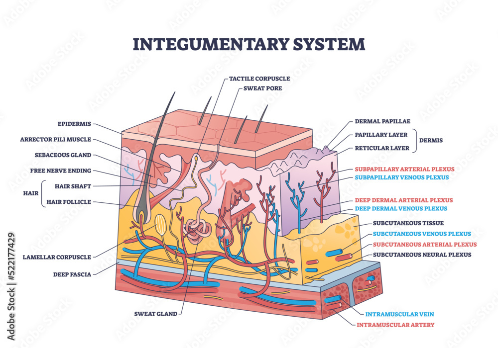
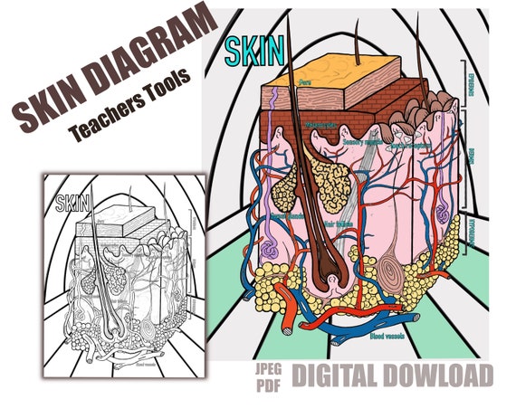
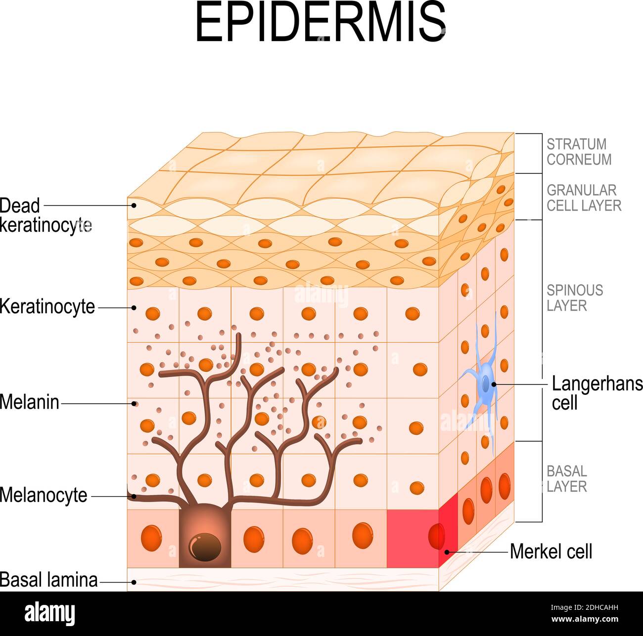
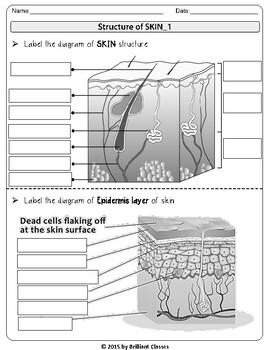
38 epidermis diagram labeled
Anatomy of the Epidermis with Pictures - Verywell Health Web20 nov. 2022 · The epidermis is the uppermost layer of your skin. It is responsible for creating skin tone and protecting against toxins and infection . Within the epidermis, there … Structure of the epidermis - DermNet NZ WebThe epidermis has a complex structure designed to protect from the environment. It has an undulating surface with cross-crossing ridges and valleys, with invaginations due to follicles and sweat duct ostia. Epidermis …
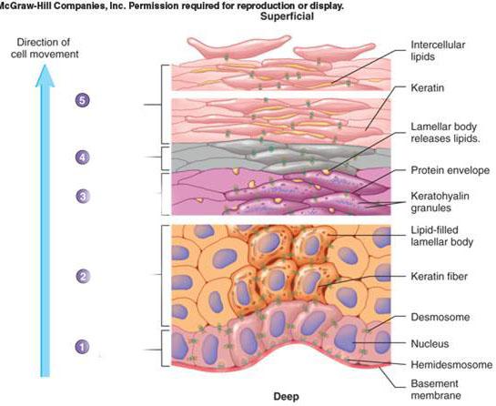
5.1 Layers of the Skin - Anatomy and Physiology 2e - OpenStax WebThe epidermis is composed of keratinized, stratified squamous epithelium. It is made of four or five layers of epithelial cells, depending on its location in the body. It does not have any …
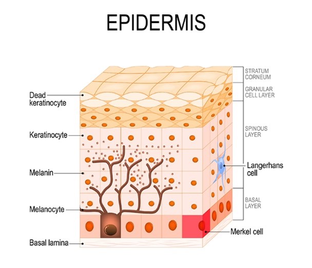
Epidermis diagram labeled
Skin Layers Diagram Images - Adobe Stock Search from thousands of royalty-free Skin Layers Diagram stock images and ... 2,076 results for skin layers diagram in all ... Labeled educational and. Epidermis (Outer Layer of Skin): Layers, Function & Structure The epidermis and the dermis are the top two layers of skin in your body. The epidermis is the top layer, and the dermis is the middle layer. The dermis exists between the epidermis and the hypodermis. While the epidermis is the thinnest layer of skin, the dermis is the thickest layer of skin. Layers of the Skin | Anatomy and Physiology I - Lumen Learning The skin is composed of two main layers: the epidermis, made of closely packed epithelial cells, and the dermis, made of dense, irregular connective tissue that ...
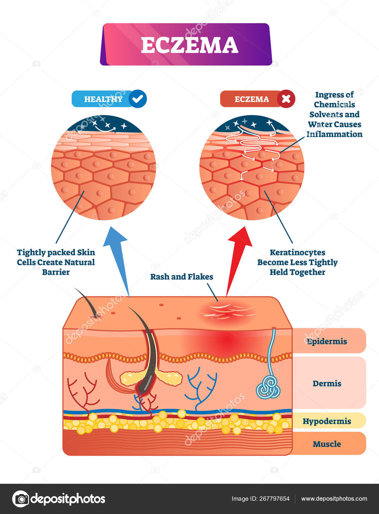
Epidermis diagram labeled. Skin 1: the structure and functions of the skin - Nursing Times Nov 25, 2019 ... The epidermis is composed mainly of keratinocytes. Beneath the epidermis is the basement membrane (also known as the dermo-epidermal junction); ... Skin Diagram Labeling Skin Diagram Labeling. 1. Label the diagram with the letters below according to the structure/area they describe. You may label with a line or put the label ... INTEGUMENTARY SYSTEM PART I: FUNCTIONS & EPIDERMIS WebThe Dermis •Is located between epidermis and subcutaneous layer •Anchors epidermal accessory structures (hair follicles, sweat glands) •Has 2 layers: –outer papillary layer … Epidermis (Outer Layer of Skin): Layers, Function & Structure WebThe epidermis is the top layer of skin in your body. It has many important functions, including protecting your body from the outside world, keeping your skin hydrated, …
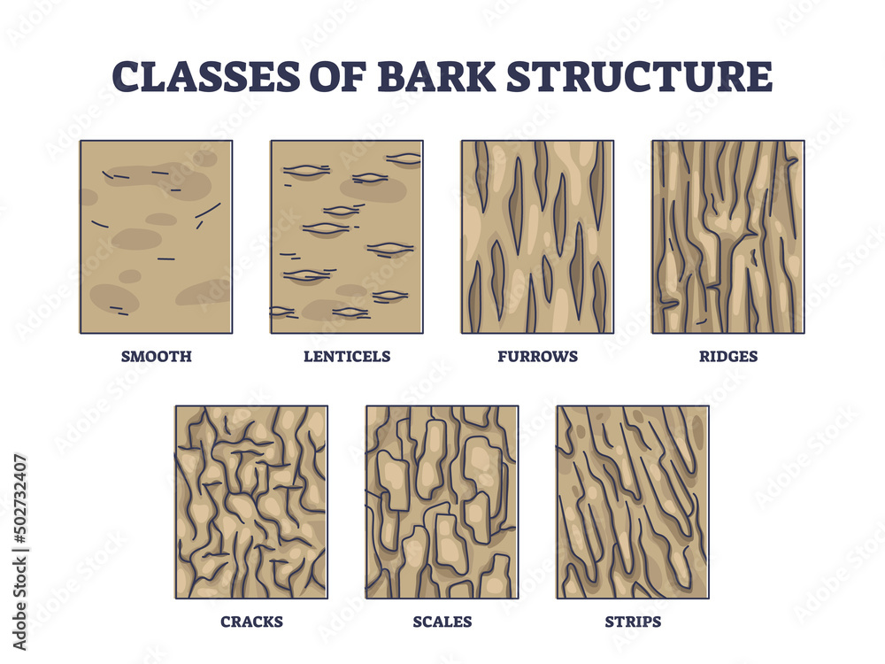
Skin Diagram Pictures, Images and Stock Photos - iStock UVB rays penetrate into epidermis of skin layer and UVA deep into the dermis. Illustration about health care and medical diagram. Structure of the epidermis - DermNet NZ Skin is made up of: Epidermis Basement membrane zone Dermis Subcutaneous tissue. Normal skin Diagram showing structural Haematoxylin and eosin stained These layers are modified according to the needs of the specific area of the body. Alila Medical Media | Human skin anatomy, labeled diagram. Human skin anatomy, hair follicle structure, sweat and sebum glands, collagen fibers, sensory and motor nerves. - Alila Medical Media. 4445 Epidermis Diagram Images, Stock Photos & Vectors Classes of bark structure with biological visual division outline diagram. Labeled educational wood epidermis classification for trees vector illustration.
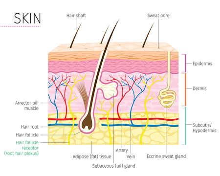
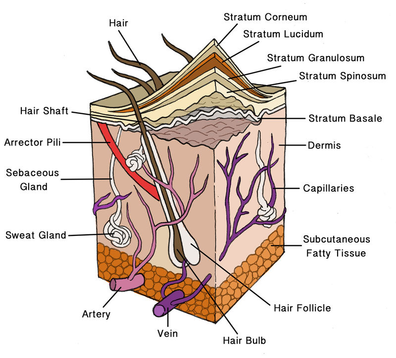
Integumentary system: Definition, diagram and function | Kenhub Oct 24, 2022 · The skin is anatomically organized as follows, from superficial to deeper layers: Epidermis Stratum basale Stratum spinosum Stratum granulosum Stratum lucidum Stratum corneum (Memorise these layers with the mnemonic: " B ritish and S panish G rannies L ove C ornflakes", see video below) Dermis Papillary dermis Reticular dermis 5.1 Layers of the Skin – Anatomy & Physiology WebFigure 5.1.1 – Layers of Skin: The skin is composed of two main layers: the epidermis, made of closely packed epithelial cells, and the dermis, made of dense, irregular connective tissue that houses blood vessels, hair … Anatomy of the Skin - Stanford Medicine Children's Health The skin is made up of 3 layers. Each layer has certain functions: Epidermis. Dermis. Subcutaneous fat layer (hypodermis) ... Integumentary system parts: Quizzes and diagrams Web14 sept. 2022 · One of the best ways to start learning about a new system, organ or region is with a labeled diagram showing you all of the main structures found within it. Not only will this introduce you to several new …
Layers of Epidermis (labeling) Diagram | Quizlet Webproduce the pigment melanin; located in deepest layer of epidermis; protection from UV radiation. Location. Term. Stratum basale. Definition. deepest epidermal layer; one layer …
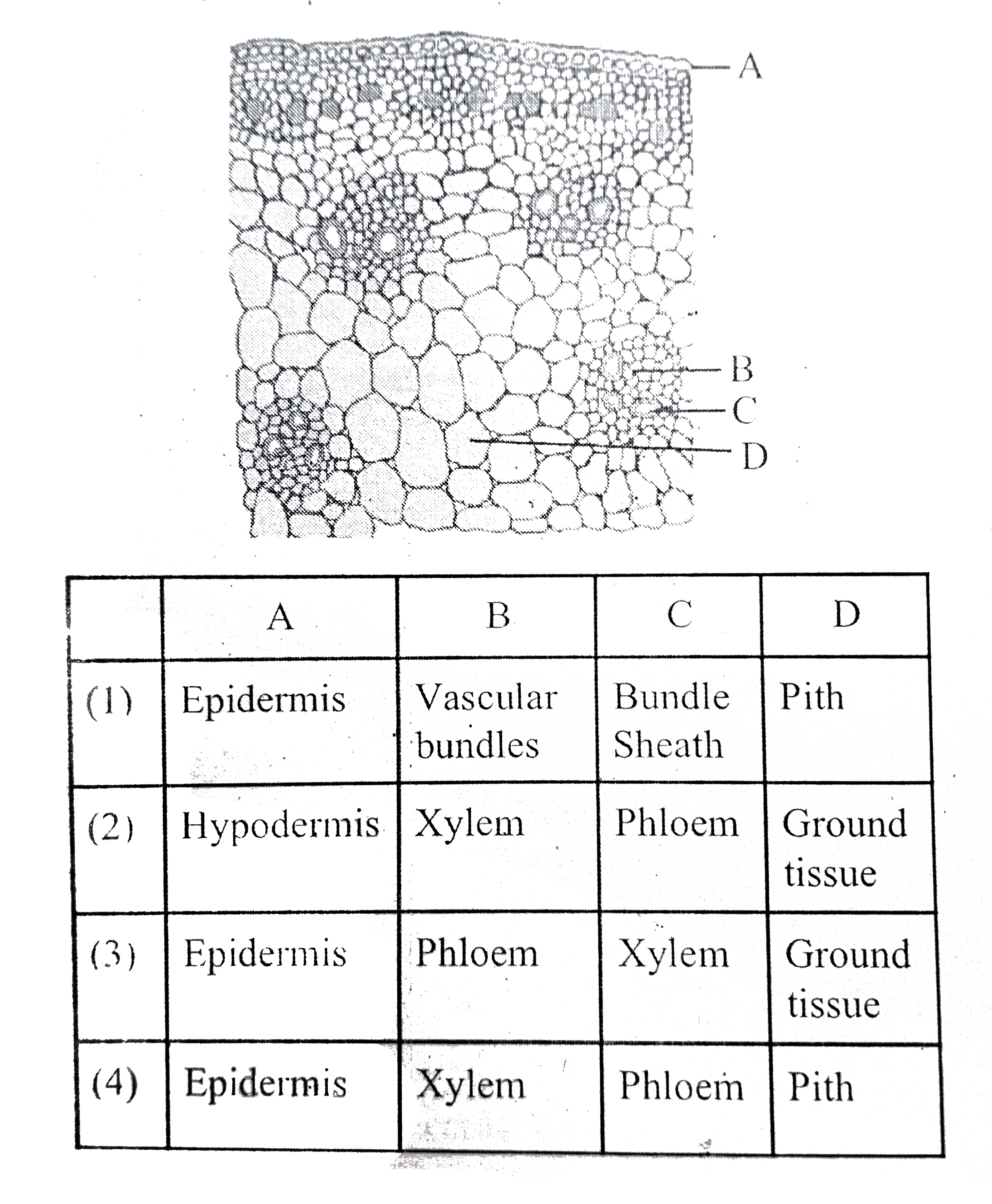
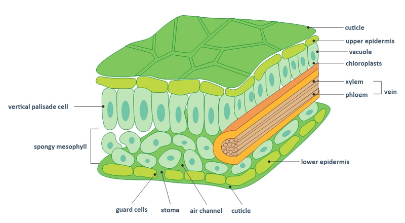
12.2: Internal Leaf Structure - Biology LibreTexts Web4 mai 2022 · The upper epidermis is a single layer of parenchyma cells. There are no stomata present in the upper epidermis of this leaf. Below the epidermis, cells (appearing pink due to staining of the nuclei and …
5.1 Layers of the Skin – Anatomy & Physiology The epidermis is composed of keratinized, stratified squamous epithelium. It is made of four or five layers of epithelial cells, depending on its location in the body. It does not have any blood vessels within it (i.e., it is avascular). Skin that has four layers of cells is referred to as “thin skin.”
Epidermis: anatomy, structure, cells and function. | Kenhub Dec 5, 2022 · The epidermis is the most superficial layer of the skin. The other two layers beneath the epidermis are the dermis and hypodermis. The epidermis is also comprised of several layers including the stratum basale, stratum spisosum, stratum granulosum, stratum lucidum, and stratum corneum.
Anatomy of the Epidermis with Pictures - Verywell Health
Skin Diagram || How to draw and label the parts of skin - Pinterest Nov 10, 2022 - 'Skin Diagram || How to draw and label the parts of skin' is demonstrated in this video tutorial step by step.The sense of touch had received ...
Layers of the Skin | Anatomy and Physiology I - Lumen Learning The skin is composed of two main layers: the epidermis, made of closely packed epithelial cells, and the dermis, made of dense, irregular connective tissue that ...
Epidermis (Outer Layer of Skin): Layers, Function & Structure The epidermis and the dermis are the top two layers of skin in your body. The epidermis is the top layer, and the dermis is the middle layer. The dermis exists between the epidermis and the hypodermis. While the epidermis is the thinnest layer of skin, the dermis is the thickest layer of skin.
Skin Layers Diagram Images - Adobe Stock Search from thousands of royalty-free Skin Layers Diagram stock images and ... 2,076 results for skin layers diagram in all ... Labeled educational and.
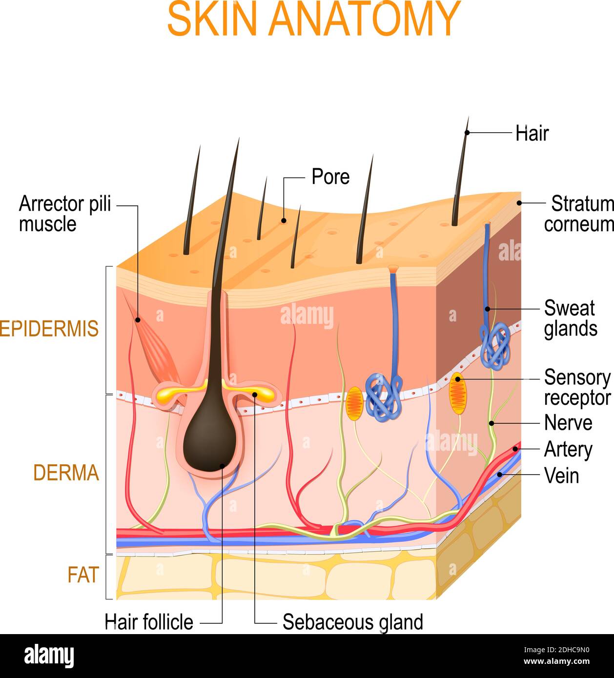
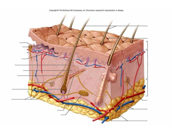


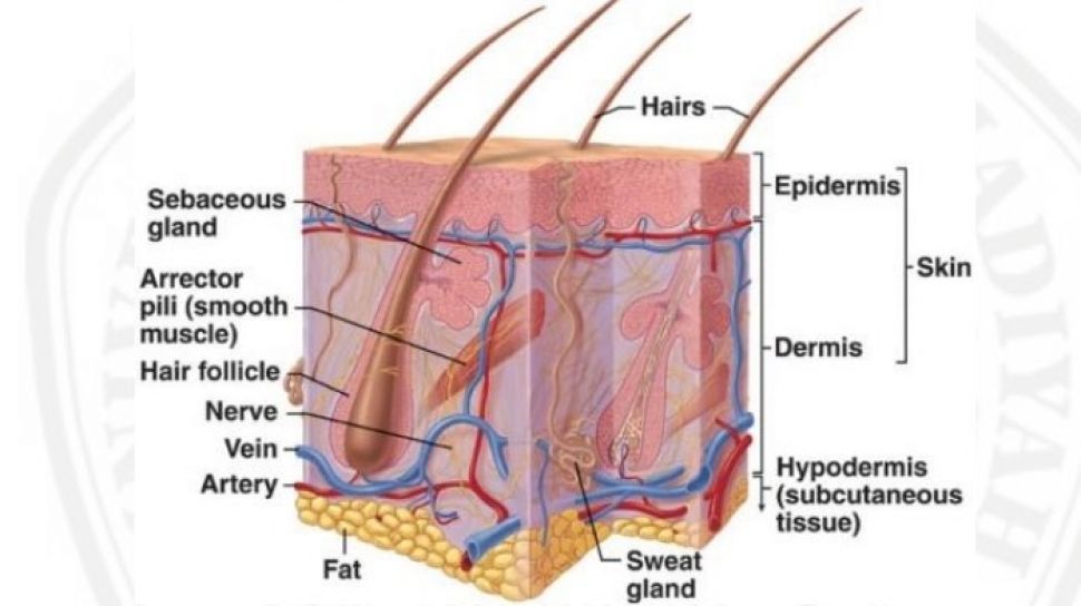












![BIOCHEMISTRY / CROSS SECTION OF A LEAF [BASIC] - Pathwayz](https://www.pathwayz.org/Node/Image/url/aHR0cHM6Ly9pLmltZ3VyLmNvbS94bUZEUXhNLnBuZz8x)
Post a Comment for "38 epidermis diagram labeled"