44 draw the heart and label the parts
How to Draw the Internal Structure of the Heart (with Pictures) - wikiHow To draw the internal structure of a human heart, follow the steps below. Part 1 Finding a Diagram 1 To find a good diagram, go to Google Images, and type in "The Internal Structure of the Human Heart". Find an image that displays the entire heart, and click on it to enlarge it. [1] 2 Find a piece of paper and something to draw with. artsexperiments.withgoogle.com › draw-to-artDraw to Art - Experiments with Google Draw with shapes on the left to discover matching artworks on the right. Use the thumbnails along the bottom to browse your matches. How does it work? We used the Sketchy dataset to match doodles to paintings, sculptures and drawings from Google Arts and Culture partner's collections around the world. Credits: Draw to Art was created by
Labelling the heart — Science Learning Hub Labelling the heart. The heart is a muscular organ that pumps blood through the blood vessels of the circulatory system. Blood transports oxygen and nutrients to the body. It is also involved in the removal of metabolic wastes. In this activity, students use online and paper resources to identify and label the main parts of the heart.
Draw the heart and label the parts
draw and label the heart draw and label the heart Human Body: Lesson Six: Muscular System (Part Two). 16 Images about Human Body: Lesson Six: Muscular System (Part Two) : Heart And Labels Drawing at GetDrawings | Free download, Circulatory System Review Guide and also heart diagram unlabeled - Google Search | A&P | Pinterest | Heart. drawisland.comDrawing tool Draw, create shape, save your online drawings with this website. You can draw online : change sizes, colors and use shapes like rectangle, round,.... and save result. You can easily save image (the drawing) to your computer. Import image to this Drawing tool (Max File Size : 1 Mb = 1000 Kb) CBSE Class 10 Answered - TopperLearning CBSE Class 10 Answered (a) Draw the diagram of human heart and label the following parts which: (i) Receives deoxygenated blood from vena cava. (ii) Send deoxygenated blood to lung through pulmonary artery. (iii) Receives oxygenated blood from lungs. (iv) Sends oxygenated blood to all parts of the body through aorta.
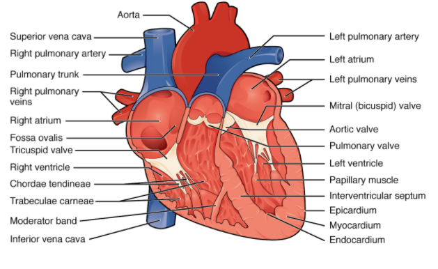
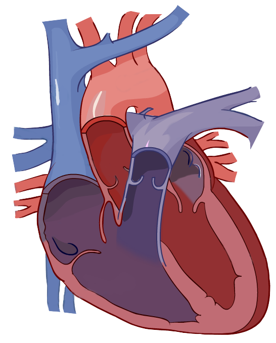
Draw the heart and label the parts. world-draw.appspot.comWorld Draw An AI Experiment to draw the world together. Let them draw the heart and label its parts let them Let them draw the heart and label its parts let them. School Rizal Technological University; Course Title BIOLOGY 01; Uploaded By ChefCaterpillarPerson352. Pages 42 Course Hero uses AI to attempt to automatically extract content from documents to surface to you and others so you can study better, e.g., in search results, to enrich docs, and more. A Labeled Diagram of the Human Heart You Really Need to See The human heart, comprises four chambers: right atrium, left atrium, right ventricle and left ventricle. The two upper chambers are called the left and the right atria, and the two lower chambers are known as the left and the right ventricles. The two atria and ventricles are separated from each other by a muscle wall called 'septum'. draw and label the heart 28 Collection Of Drawing Of A Human Heart And Its Parts - Simple Heart. 16 Pictures about 28 Collection Of Drawing Of A Human Heart And Its Parts - Simple Heart : Heart And Labels Drawing at GetDrawings | Free download, Circulatory System Review Guide and also Circulatory System Review Guide.
File:Diagram of the human heart (cropped).svg - Wikipedia Athletic heart syndrome Atrium (heart) Blood Cavoatrial junction Fourth heart sound Heart Human body Inferior vena cava Lutembacher's syndrome Mitral valve Mitral valve repair Pressure-volume diagram Pulmonary artery Pulmonary insufficiency Pulmonary valve Pulmonary vein Ross procedure Sano shunt Third heart sound Tissue engineering of heart valves Label the Heart Diagram | Quizlet Label the Heart Diagram | Quizlet Label the Heart 4.5 (53 reviews) + − Learn Test Match Created by bluesas9 Terms in this set (15) Superior Vena Cava ... Right Ventricle ... Left Atrium ... Atrioventricular/Tricuspid Valve ... Atrioventricular/Mitral Valve ... Septum ... Right Atrium ... Semi-lunar Valves ... Left Pulmonary Veins ... Draw the sectional view of the human heart and label it. Click here👆to get an answer to your question ️ Draw the sectional view of the human heart and label it. Solve Study Textbooks Guides. Join / Login >> Class 10 >> Biology ... >> Draw the sectional view of the human hea. Question . Draw the sectional view of the human heart and label it. ... › dictionary › drawDraw Definition & Meaning - Merriam-Webster Synonyms of draw transitive verb 1 : to cause to move continuously toward or after a force applied in advance : pull draw your chair up by the fire : such as a : to move (something, such as a covering) over or to one side draw the drapes b : to pull up or out of a receptacle or place where seated or carried draw water from the well drew a gun
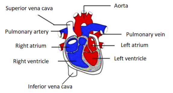
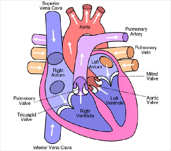
Human Heart Diagram Labeled - Science Trends Human Heart Diagram Labeled Daniel Nelson 1, January 2019 | Last Updated: 3, March 2020 The human heart is an organ responsible for pumping blood through the body, moving the blood (which carries valuable oxygen) to all the tissues in the body. Without the heart, the tissues couldn't get the oxygen they need and would die. quickdraw.withgoogle.com › dataQuick, Draw! The Data Over 15 million players have contributed millions of drawings playing Quick, Draw! These doodles are a unique data set that can help developers train new neural networks, help researchers see patterns in how people around the world draw, and help artists create things we haven’t begun to think of. › Draw4 Ways to Draw - wikiHow Nov 10, 2022 · Draw a grid on the paper if you need help with proportions. If you're drawing something from a source image, draw several evenly-spaced vertical and horizontal lines on your paper to make a grid. Then, draw the same lines on your source image. Look at each square on the source image and copy it into the corresponding square on your paper. PDF Arrows show the path of blood flow in the human heart. The blood enters ... Arrows show the path of blood flow in the human heart. The blood enters the heart from the body through the superior vena cava and the inferior vena cava. Then the blood enters the right atrium chamber of the heart. The blood then moves through the tricuspid valve (shown as two white flaps) into the right ventricle chamber of the heart.
Diagram of Human Heart and Blood Circulation in It Three veins of the heart are pulmonary vein, Venae Cavae, and coronary sinus. Pulmonary vein transfer oxygenated blood to the left side of your heart, venae cavae takes deoxygenated blood back to the heart, and coronary sinus receives deoxygenated blood and transfers it to the right atria. The Circulation of Blood
Draw a diagram of the front view of human heart and label any six parts ... Draw a diagram of the front view of human heart and label any six parts (including at least two) that are concerned with arterial blood supply to the heart muscles. Solution Coronary circulation: The circulation involving the movement of oxygenated blood towards the heart muscle is called coronary circulation.
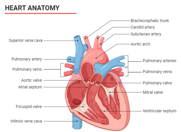
Diagrams, quizzes and worksheets of the heart | Kenhub Labeled heart diagrams Take a look at our labeled heart diagrams (see below) to get an overview of all of the parts of the heart. Once you're feeling confident, you can test yourself using the unlabeled diagrams of the parts of the heart below. Labeled heart diagram showing the heart from anterior Unlabeled heart diagrams (free download!)
PDF Name: Heart Anatomy Coloring - Weebly The heart pumps blood to the lungs and other parts of the body. It is made of cardiac muscle tissue. Cardiac muscle is a special type of muscle that does not fatigue easily. This allows the heart to continuously contract. When the heart contracts, blood is forced out of the heart. When it relaxes, the chambers of the heart fill with blood.
› discover › learning-to-drawHow to draw for beginners | Learn to draw | Adobe Learn to draw better by copying. Building off the work of those who’ve come before you is a great way to learn. Trying to pass off the work of another artist as your own is plagiarism, but emulating the work of accomplished illustrators is an observational exercise that can help you improve your drawing skills.
Heart Anatomy: Labeled Diagram, Structures, Blood Flow ... - EZmed We now have a 2x2 table in which we can label the boxes/chambers of the heart. Box 1: The first box is located in the right upper region. We know the atria are on top, and since box 1 is located on the right side, this is the right atrium. Box 2: The second box is also located on the right side, but now we are in the lower region.
Solved draw the human heart and label all the parts and give | Chegg.com Human heart is located between the lungs, slightly tilted towards the left side of sternum. The function of heart is to pump the blood and make it circulate constantly throughout the body. Here are functions of some important part of the heart. 1. At ….
Heart Diagram with Labels and Detailed Explanation - BYJUS Well-Labelled Diagram of Heart The heart is made up of four chambers: The upper two chambers of the heart are called auricles. The lower two chambers of the heart are called ventricles. The heart wall is made up of three layers: The outer layer of the heart wall is called epicardium. The middle layer of the heart wall is called myocardium.
Label the heart — Science Learning Hub In this interactive, you can label parts of the human heart. Drag and drop the text labels onto the boxes next to the heart diagram. If you want to redo an answer, click on the box and the answer will go back to the top so you can move it to another box. If you want to check your answers, use the Reset Incorrect button.
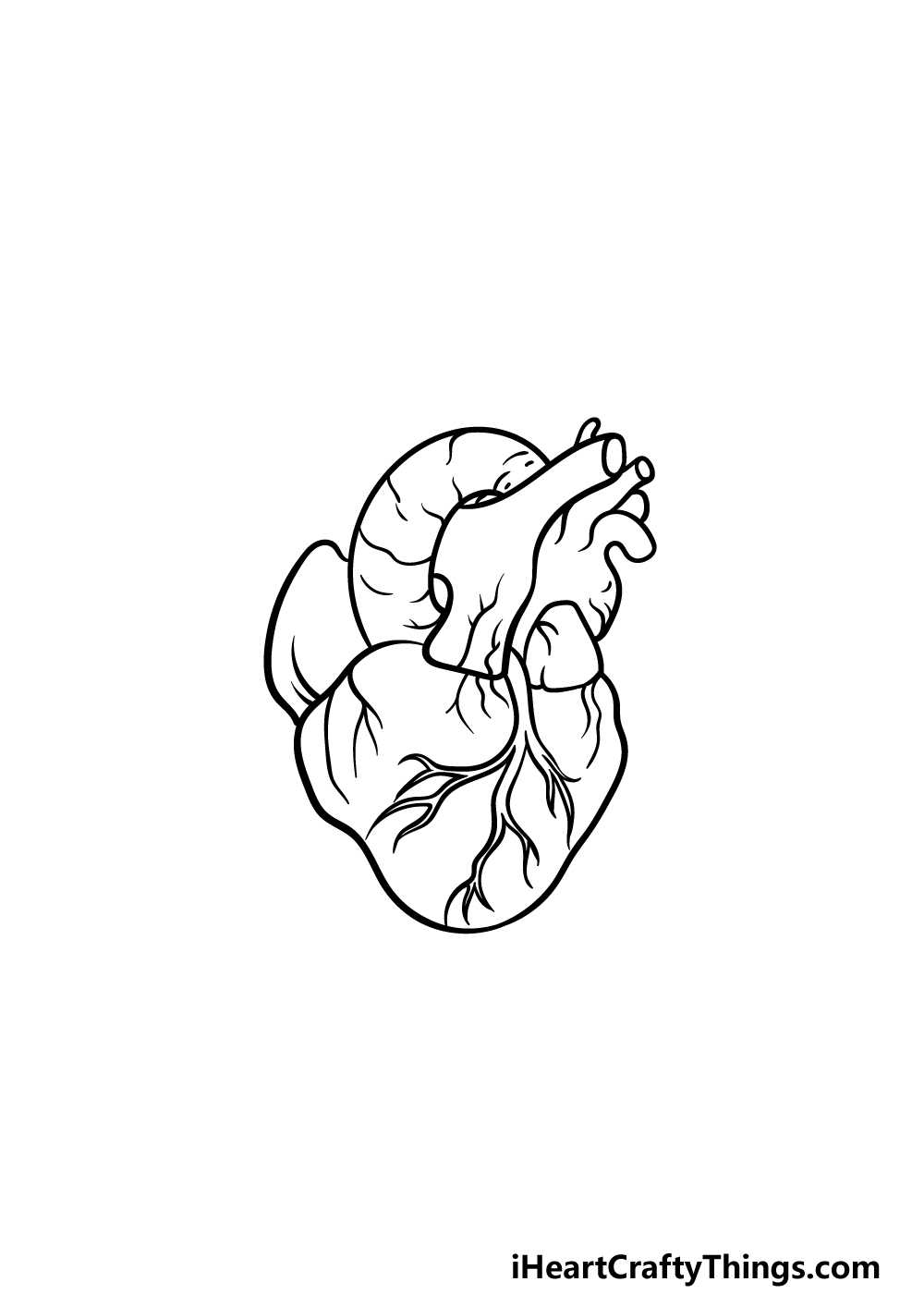
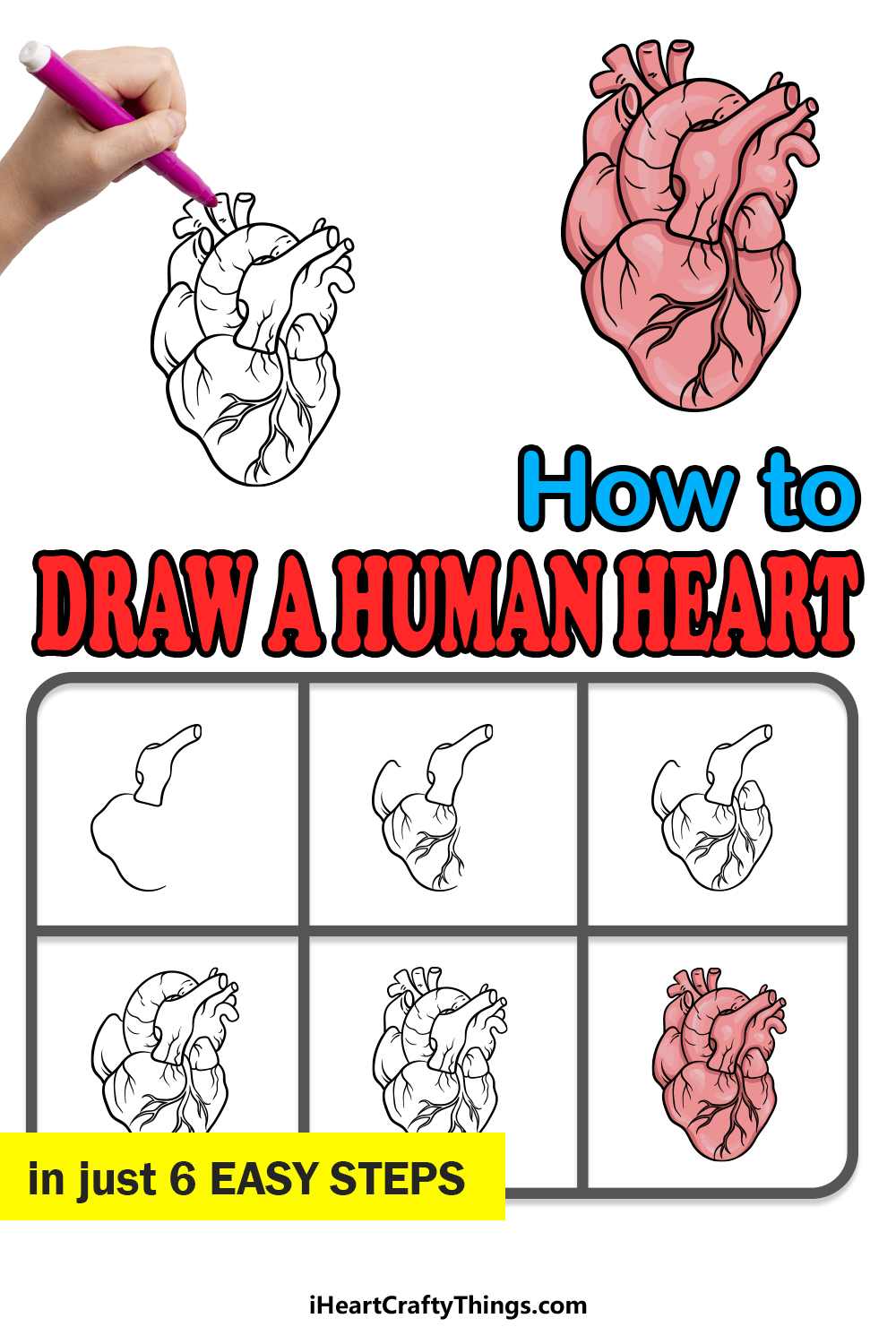
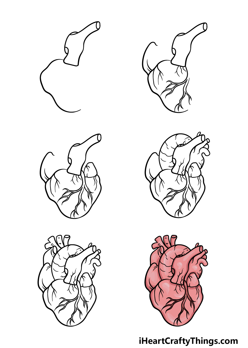
Easy way to draw heart structure by 5 steps | labeling of heart ... Easy way to draw heart structure by 5 steps .... 1) a basic structure , 2) a T shape structure 3) a opposite question mark 4) a straight line structure 5) 6 V shape structure
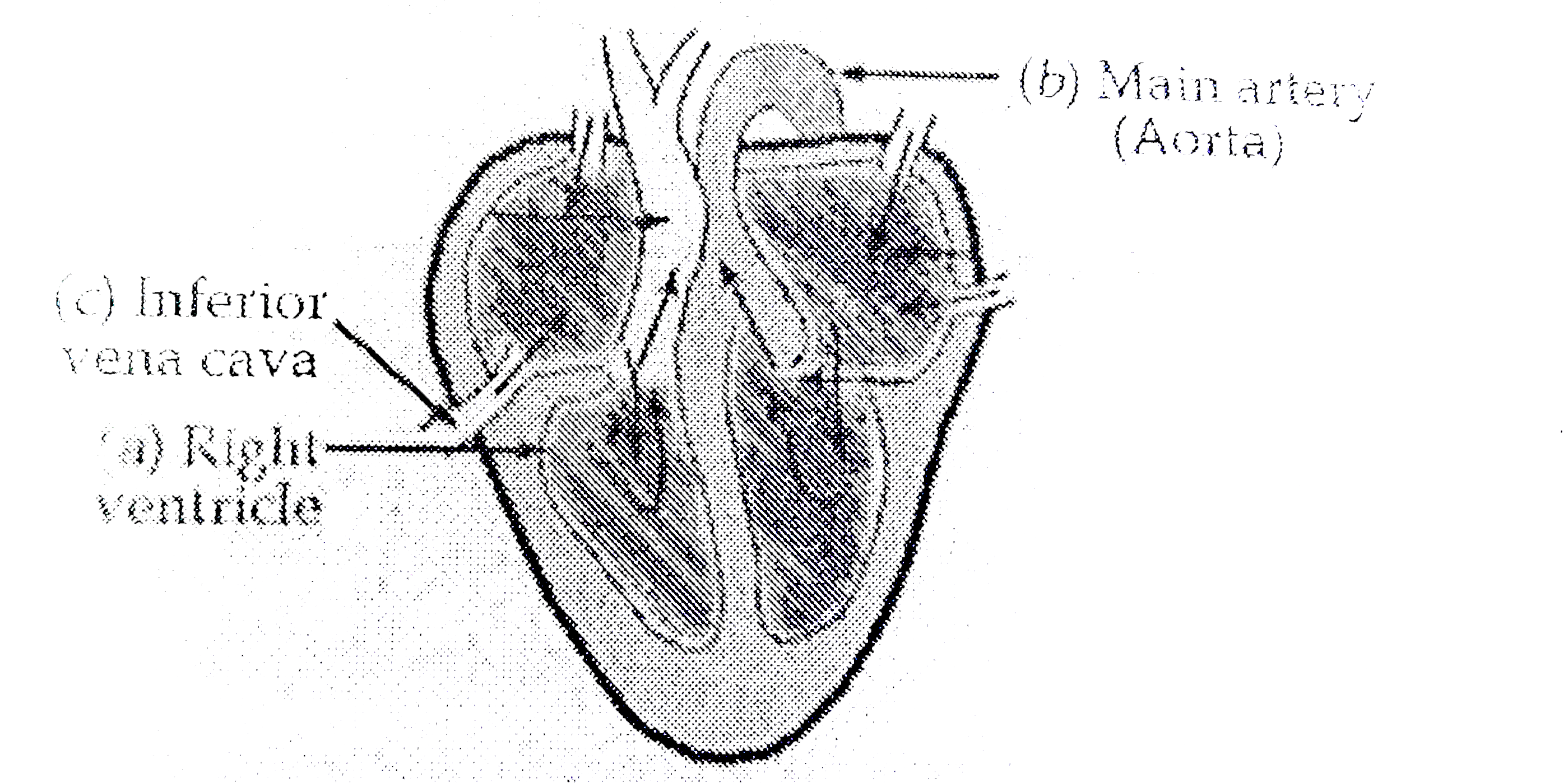
Academic Articles Updated on 10-Oct-2022 10:17:18. (a) The human circulatory system is responsible for the transport of materials inside the human body. The organs of the circulatory system are the heart, arteries, veins, and capillaries. (b) Given below is an image of the human heart with its main parts.

How to draw Human Heart with colour | Human Heart labelled diagram|Human Heart drawing easy tutorial
Easy way to draw human heart and label its main parts Easy way to draw human heart and label its main parts 307 views Dec 28, 2018 44 Dislike Share Save Amal Sunder A 1.93K subscribers Hai friends, In this video I am drawing a human heart...
A Diagram of the Heart and Its Functioning Explained in Detail Heart pumps pure blood to different parts of the body and then takes the deoxygenated blood from all the parts to the lungs for oxygenation. Normally in a minute the heart beats 72 times and pumps around 1,500 to 2,000 gallons of blood per day. Let's check out heart diagram which can help you to understand functioning of the heart in a better way.
Cross Section of the Heart Diagram & Function | Body Maps - Healthline Each of the four chambers of the heart has its own valve. They are: Tricuspid valve: This valve is located between the right atrium and right ventricle. It is also called the right AV valve....
Label Parts Of A Heart - Label The Heart Diagram Quizlet Walt label parts of the heart. The main artery carrying oxygenated blood to all parts of the body · pulmonary artery: The heart is a muscular organ about the size of a fist, located just behind and slightly left of the breastbone. The human heart right atrium tricuspid valve right .
Conducting System of Human Heart (With Diagram) The velocity of conduction of the impulse in the different parts of human heart is: i. SA and AV nodes—0.05 m/sec. ii. Atrial and ventricular muscles—1.0 m/sec. ADVERTISEMENTS: iii. Internodal fibers, Bundle of His and its branches— 1.0 m/sec.
Picture of the Heart - WebMD The heart is a muscular organ about the size of a fist, located just behind and slightly left of the breastbone. The heart pumps blood through the network of arteries and veins called the ...
CBSE Class 10 Answered - TopperLearning CBSE Class 10 Answered (a) Draw the diagram of human heart and label the following parts which: (i) Receives deoxygenated blood from vena cava. (ii) Send deoxygenated blood to lung through pulmonary artery. (iii) Receives oxygenated blood from lungs. (iv) Sends oxygenated blood to all parts of the body through aorta.
drawisland.comDrawing tool Draw, create shape, save your online drawings with this website. You can draw online : change sizes, colors and use shapes like rectangle, round,.... and save result. You can easily save image (the drawing) to your computer. Import image to this Drawing tool (Max File Size : 1 Mb = 1000 Kb)
draw and label the heart draw and label the heart Human Body: Lesson Six: Muscular System (Part Two). 16 Images about Human Body: Lesson Six: Muscular System (Part Two) : Heart And Labels Drawing at GetDrawings | Free download, Circulatory System Review Guide and also heart diagram unlabeled - Google Search | A&P | Pinterest | Heart.
















Post a Comment for "44 draw the heart and label the parts"