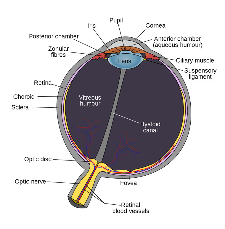
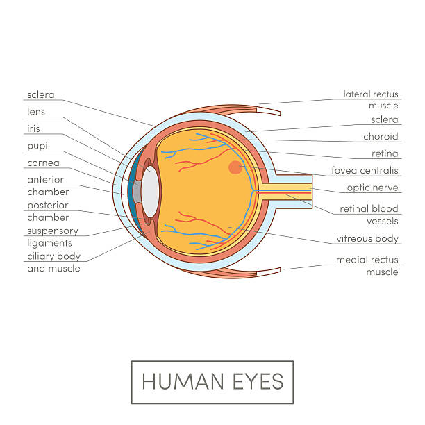
42 human eye labeled diagram
› c › huMANs_channelhuMAN - YouTube Consider that EVERYTHING in your life is a relationship. From your relationship to yourself, others, the truth, and even things and hobbies. ... everything. Let's be BLUNT about the hypocrisy and ... Human eye | Definition, Anatomy, Diagram, Function, & Facts the lid may be divided into four layers: (1) the skin, containing glands that open onto the surface of the lid margin, and the eyelashes; (2) a muscular layer containing principally the orbicularis oculi muscle, responsible for lid closure; (3) a fibrous layer that gives the lid its mechanical stability, its principal portions being the tarsal …
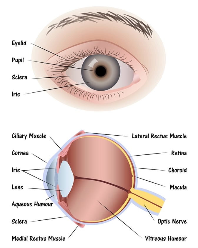
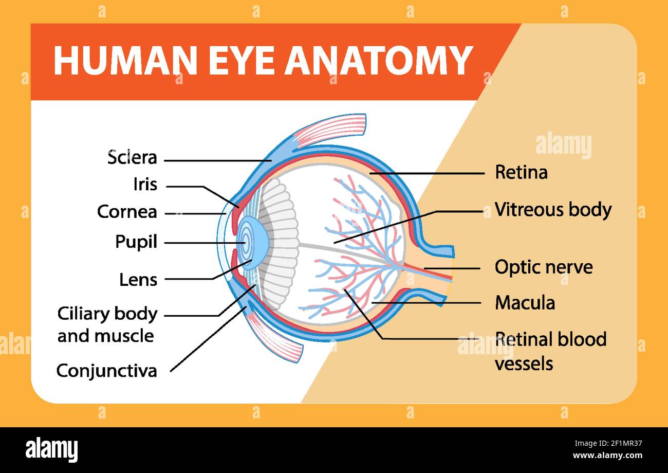
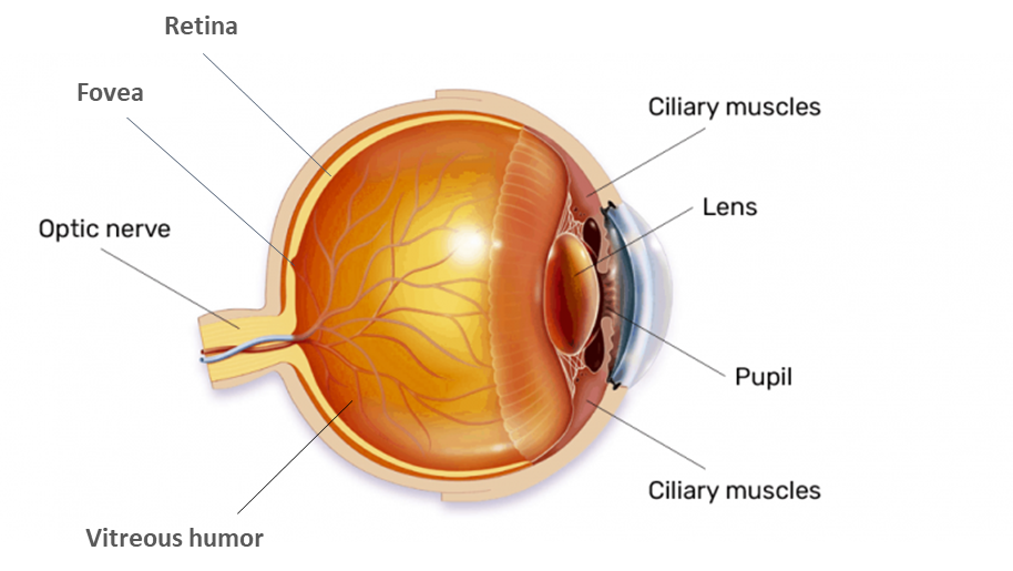
What Does the Eye Look Like? - Diagram of the Eye | Harvard Eye Associates The eye is one of the most complicated organs in the human body. Major parts of the eye include the cornea, pupil, lens, retina and macula. Starting from "front" to "back" of the eye, the cornea is in charge of shaping the light as it comes into the eye and passes through the lens.

Human eye labeled diagram
› search › human14,158,752 Human Images, Stock Photos & Vectors | Shutterstock Find Human stock images in HD and millions of other royalty-free stock photos, illustrations and vectors in the Shutterstock collection. Thousands of new, high-quality pictures added every day. Human Eye Ball Anatomy & Physiology Diagram - eMedicineHealth Human Eye Ball Anatomy & Physiology Diagram Anatomy and Physiology of the Eye Facts & Orbit Eyes & Eyelashes Conjunctiva & Sclera Chambers Cornea, Iris, and Pupil Lens, Vitreous Cavity Retina/Macula/Choroid Optic Nerve Muscles Guide Eye Anatomy, Function, and Physiology Facts Parts of the eye. Labeled Eye Diagram - Science Trends The human eye is composed of many different parts that work together to interpret the world around us. What you want to interpret as a major part of the human eye is somewhat up to the individual, but in general there are seven parts of the human eye: the cornea, the pupil, the iris, the ... Labeled Eye Diagram. Daniel Nelson PRO INVESTOR. 24 ...
Human eye labeled diagram. Human Muscle Diagram Pictures, Images and Stock Photos Browse 3,338 human muscle diagram stock photos and images available, or start a new search to explore more stock photos and images. Sort by: Most popular. Male fitness model. Male muscular anatomy vector scheme - posterior and anterior view. Fitness training, muscles street workout. james-camerons-avatar.fandom.com › wiki › HumanHuman | Avatar Wiki | Fandom Humans (Na'vi name: tawtute meaning "sky people") are a sapient, sentient, bipedal mammalian dominant species native to planet Earth, who by the 22nd century has become a technologically advanced species capable of interstellar travel and colonization. Humanity is the first and only alien species known to the Na'vi, another sapient species native to Pandora, where the humans arrived in search ... diagram of human eye with labelling Eye diagram anatomy labeled labels ... We have 9 Images about Diagram of the human eye - Primary KS2 teaching resource - Scholastic like Human Eye - Discovering DNA, Eye Anatomy Diagram - EnchantedLearning.com and also Eye Anatomy Diagram - EnchantedLearning.com. Read more: Diagram Of The Human Eye - Primary KS2 Teaching Resource - Scholastic education.scholastic.co.uk Eye Anatomy: 16 Parts of the Eye & Their Functions - Vision Center The following are parts of the human eyes and their functions: 1. Conjunctiva The conjunctiva is the membrane covering the sclera (white portion of your eye). The conjunctiva also covers the interior of your eyelids. Conjunctivitis, often known as pink eye, occurs when this thin membrane becomes inflamed or swollen.
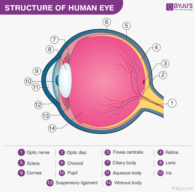
Eye Pictures, Anatomy & Diagram | Body Maps - Healthline Eyes are approximately one inch in diameter. Pads of fat and the surrounding bones of the skull protect them. The eye has several major components: the cornea, pupil, lens, iris, retina, and... Structure and Functions of Human Eye with labelled Diagram - BYJUS Structure of the Eye. The eye is one of the sensory organs of the body. In this article, we shall explore the anatomy of the eye. The structure of the eye is an important topic to understand as it one of the important sensory organs in the human body. It is mainly responsible for vision, differentiation of colour (the human eye can differentiate approximately 10 - 12 million colours) and maintaining the biological clock of the human body. Labelling the eye — Science Learning Hub The human eye has several structures that enable entering light energy to be converted to electrochemical energy. This stimulates the visual centres in the brain, giving us the sensation of seeing. In this interactive, you can label parts of the human eye. Use your mouse or finger to hover over a box to highlight the part to be named. Eye Diagram With Labels and detailed description - BYJUS Diagram Of Eye Diagram Of Eye The human eye is responsible for the most important function of the human body, the sense of sight. It consists of several distinct parts that work in coordination with each other. The most common eye diseases include myopia, hypermetropia, glaucoma and cataract.
Diagram of the Eye - Lions Eye Institute To understand the eye and its functions, it's important to understand how the eye works, see below diagrams for both the external eye and the internal eye. The External Eye Instructions Click the parts of the eye to see a description for each. Hover the diagram to zoom. The Internal Eye Instructions Eye Anatomy: A Closer Look At the Parts of the Eye - All About Vision Human Eye Anatomy (seen from above) For more details about specific structures of the eye and how they function, visit these pages: Conjunctiva Of The Eye Sclera: The White Of The Eye Cornea Of The Eye The Uvea Of The Eye Pupil: Aperture Of The Eye The Retina: Where Vision Begins Macula Lutea Of The Eye Choroid Of The Eye Lens Of The Eye m.youtube.com › watchRag'n'Bone Man - Human (Official Video) - YouTube Rag'n'Bone Man - Human (Official Video)Stream Rag'n'Bone Man here: to Rag'n'Bone Man's YouTube Channel: https:/... › thesaurus › human73 Synonyms & Antonyms of HUMAN - Merriam-Webster Synonyms for HUMAN: natural, mortal, humanoid, hominid, humanlike, anthropoid, earthborn, creatural; Antonyms of HUMAN: nonhuman, divine, superhuman, supernatural ...
The Eyes (Human Anatomy): Diagram, Optic Nerve, Iris, Cornea ... - WebMD The Eyes (Human Anatomy): Diagram, Optic Nerve, Iris, Cornea, Pupil, & More Menu Eye Health Reference A Picture of the Eye Written by WebMD Editorial Contributors Medically Reviewed by...
m.youtube.com › watchChristina Perri - Human [Official Video] - YouTube the official music video of “human” from the album ‘head or heart’ buy/listen to 'head or heart': by elliot...
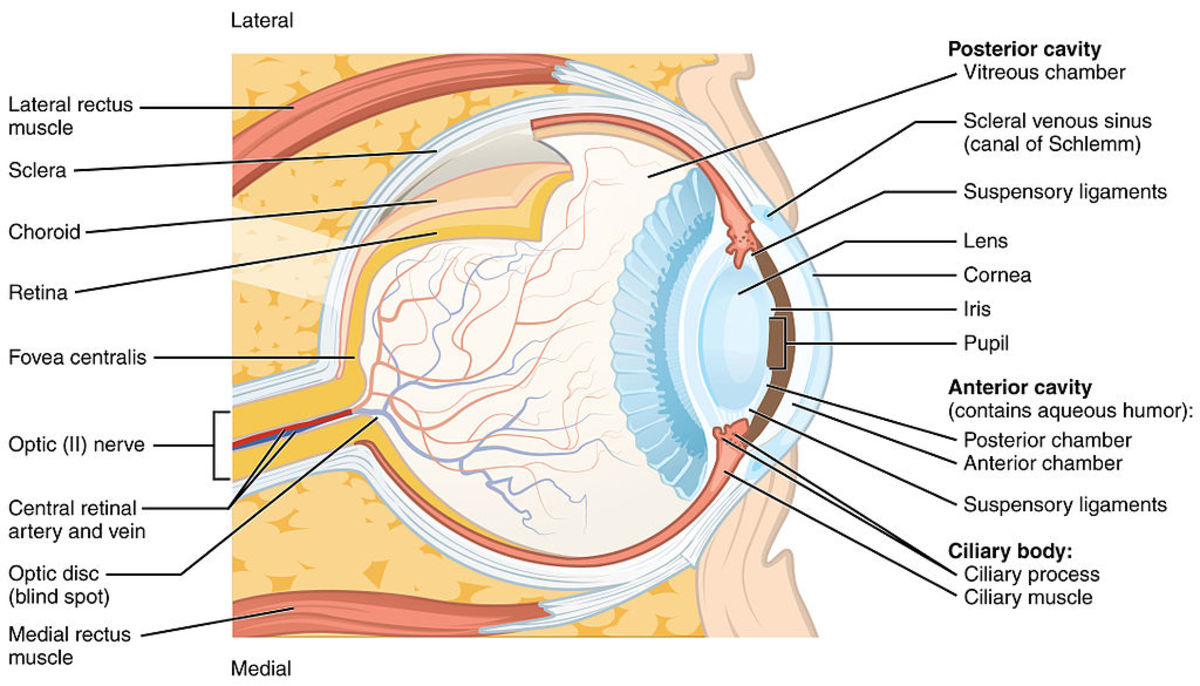
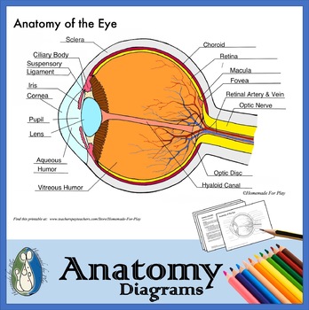
Eye Anatomy: Parts of the Eye and How We See Eye Anatomy: Parts of the Eye Outside the Eyeball. The eye sits in a protective bony socket called the orbit. Six extraocular muscles in the orbit are attached to the eye. These muscles move the eye up and down, side to side, and rotate the eye. The extraocular muscles are attached to the white part of the eye called the sclera. This is a strong layer of tissue that covers nearly the entire surface of the eyeball.
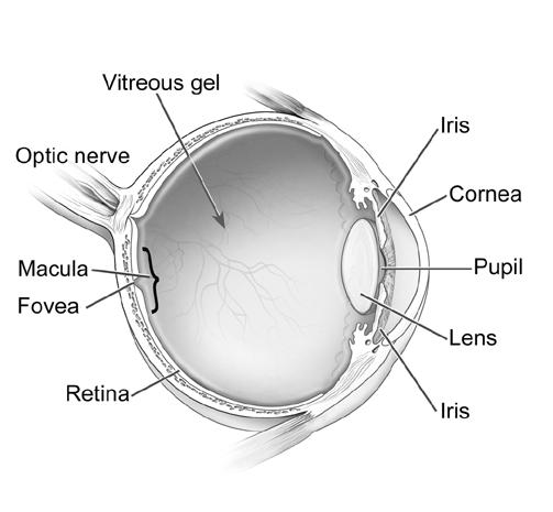
PDF Parts of the Eye - National Institutes of Health Eye Diagram Handout Author: National Eye Health Education Program of the National Eye Institute, National Institutes of Health Subject: Handout illustrating parts of the eye Keywords: parts of the eye, eye diagram, vitreous gel, iris, cornea, pupil, lens, optic nerve, macula, retina Created Date: 12/16/2011 12:39:09 PM
Human Eye Diagram, How The Eye Work -15 Amazing Facts of Eye Human Eye Diagram [pic:nei.nih.gov] What are the Three Layers of the Human Eye? The Outer Layer - The Cornea and Sclera The Middle Layer - Iris, The Choroid, and The Ciliary Body The Inner Layer :-The Retina What are all parts of the human eye and their functions? Sclera The Sclera is a strong outer white part of the eye .
6,819 Human eye diagram Images, Stock Photos & Vectors | Shutterstock Find Human eye diagram stock images in HD and millions of other royalty-free stock photos, illustrations and vectors in the Shutterstock collection. Thousands of new, high-quality pictures added every day.
Human Eye Anatomy Quiz - Sporcle Human Eye Anatomy Can you locate the parts of the human eye? By smac17. Plays. Comments. Comments. Bookmark Quiz Bookmark Quiz -/5-RATE QUIZ. YOU. MORE INFO Picture Click Click on regions of an image Click on regions of an image Forced Order Answers have to be entered in order Answers have to be entered in order ...
Eye Anatomy: Diagram & Human Eye Anatomy | StudySmarter Eye Anatomy Bioenergetics Investigating Photosynthesis Biological Molecules ATP Carbohydrates Condensation Reaction DNA and RNA DNA replication Denaturation Enzymes Factors Affecting Enzyme Activity Fatty Acids Hydrolysis Reaction Inorganic Ions Lipids Measuring enzyme-controlled reactions Monomers Monomers and Polymers Monosaccharides
Labeled Eye Diagram | Eye anatomy diagram, Eye anatomy, Diagram of the eye Human Eye Diagram Diagram Of The Eye Brain Anatomy Anatomy And Physiology Human Anatomy Anatomy Organs Body Anatomy Eyes More information ... More information Labeled Eye Diagram Comments More like this Optometry School Optometry Students Vet School Medical School Opthalmic Technician Optician Training Retinal Degeneration Eye Facts Eye Anatomy
Human Eye - Definition, Structure, Function, Parts, Diagram - BYJUS Structure of Human Eye A human eye is roughly 2.3 cm in diameter and is almost a spherical ball filled with some fluid. It consists of the following parts: Sclera: It is the outer covering, a protective tough white layer called the sclera (white part of the eye). Cornea: The front transparent part of the sclera is called the cornea.
Class 10 Human Eye Labelled Diagram drawing // Chapter 11The human eye ... Class 10 Human eye diagram drawing How to draw Human eye class 10 Science labelled diagram drawing class 10 chapter 11#cbse #ncert #class10
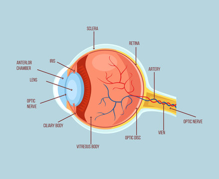
Anatomy of the Eye | Johns Hopkins Medicine Ciliary body. The part of the eye that produces aqueous humor. Cornea. The clear, dome-shaped surface that covers the front of the eye. Iris. The colored part of the eye. The iris is partly responsible for regulating the amount of light permitted to enter the eye. Lens (also called crystalline lens).
Labelled Diagram of Human Eye, Explanation and Function - VEDANTU The human eye is a part of the sensory nervous system. Labeled Diagram of Human Eye The eyes of all mammals consist of a non-image-forming photosensitive ganglion within the retina which receives light, adjusts the dimensions of the pupil, regulates the availability of melatonin hormones, and also entertains the body clock.
Draw the labelled diagram of human eye and explain the image formation ... The human eye is like a camera. Its lens system forms an image on a light-sensitive screen called the retina. Light enters the eye through a thin membrane called the cornea. It forms a transparent bulge on the front surface of the eyeball. The eyeball is approximately spherical in shape with a diameter of about 2.3 cm.
› science › human-bodyHuman body | Organs, Systems, Structure, Diagram, & Facts Dec 2, 2022 · Chemically, the human body consists mainly of water and of organic compounds —i.e., lipids, proteins, carbohydrates, and nucleic acids. Water is found in the extracellular fluids of the body (the blood plasma, the lymph, and the interstitial fluid) and within the cells themselves. It serves as a solvent without which the chemistry of life ...
The Anatomy of Human Eye with Diagram | EdrawMax Online - Edrawsoft The human eye diagram is a visual depiction of the human eye. The following aspects are essential when constructing a human eye diagram . Source: EdrawMax 1.1 Conjunctiva of the Eye The conjunctiva is a thin, translucent layer of tissue that protects the front of the eyes, including the sclera and the eyelids inner surface.
Eye Anatomy - 3D model by MotionCow [5dac474] - Sketchfab 101.2k. 89. Triangles: 64.9k. Vertices: 51.7k. More model information. Realistic cross-section of the Eye. This 3D model can be licensed from MotionCow by Educators, 3D Artists and App Developers. Published 6 years ago. Science & technology 3D Models.
Labeled Eye Diagram - Science Trends The human eye is composed of many different parts that work together to interpret the world around us. What you want to interpret as a major part of the human eye is somewhat up to the individual, but in general there are seven parts of the human eye: the cornea, the pupil, the iris, the ... Labeled Eye Diagram. Daniel Nelson PRO INVESTOR. 24 ...
Human Eye Ball Anatomy & Physiology Diagram - eMedicineHealth Human Eye Ball Anatomy & Physiology Diagram Anatomy and Physiology of the Eye Facts & Orbit Eyes & Eyelashes Conjunctiva & Sclera Chambers Cornea, Iris, and Pupil Lens, Vitreous Cavity Retina/Macula/Choroid Optic Nerve Muscles Guide Eye Anatomy, Function, and Physiology Facts Parts of the eye.
› search › human14,158,752 Human Images, Stock Photos & Vectors | Shutterstock Find Human stock images in HD and millions of other royalty-free stock photos, illustrations and vectors in the Shutterstock collection. Thousands of new, high-quality pictures added every day.

:max_bytes(150000):strip_icc()/GettyImages-695204442-b9320f82932c49bcac765167b95f4af6.jpg)













Post a Comment for "42 human eye labeled diagram"