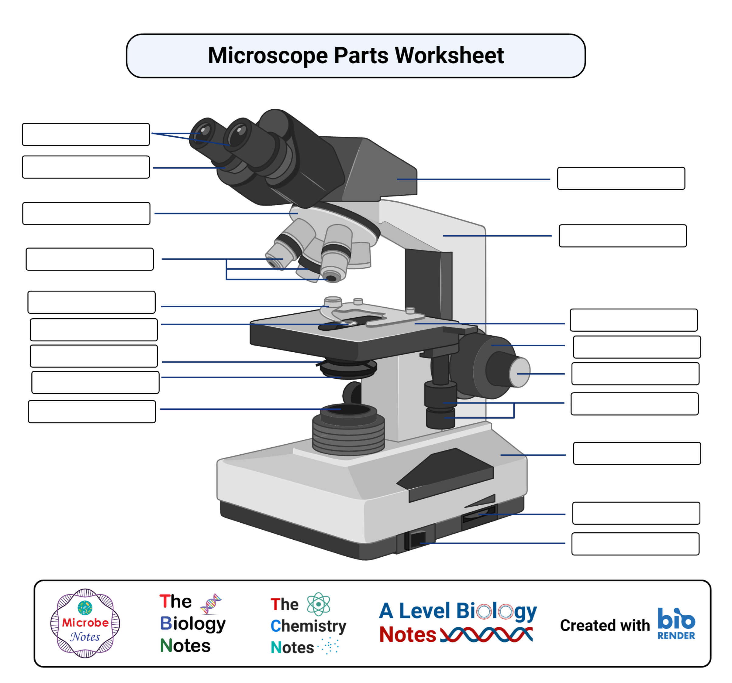
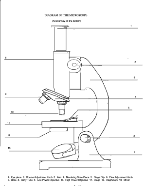
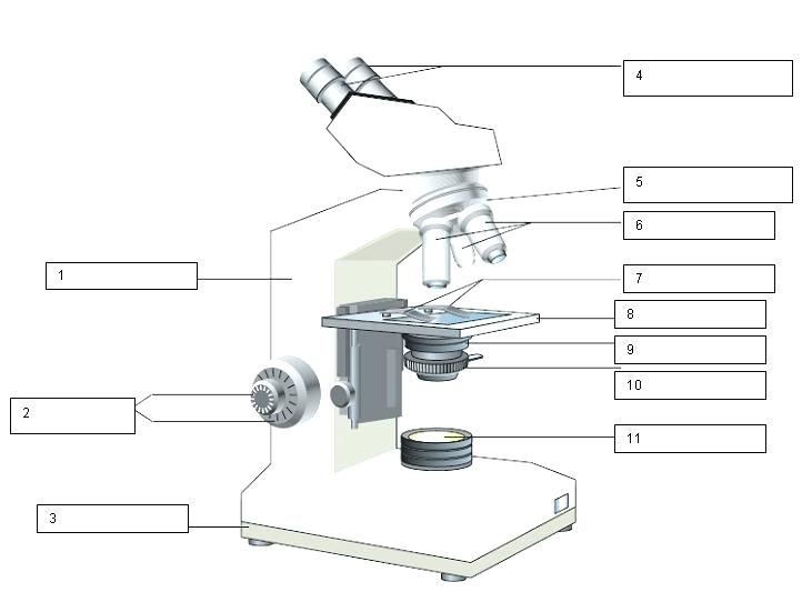
42 blank compound microscope diagram
Microscope Labeling - The Biology Corner 18. You should carry the microscope by the _____ and the _____. 19. The objectives are attached to what part of the microscope (it can be rotated to click lenses into place?) _____ 20. A microscope has an ocular objective of 10x and a high power objective of 50x, what is the microscope's total magnification? Compound Microscope Diagram Blank - 16 images - types of microscopes ... Here are a number of highest rated Compound Microscope Diagram Blank pictures on internet. We identified it from reliable source. Its submitted by presidency in the best field. We put up with this kind of Compound Microscope Diagram Blank graphic could possibly be the most trending subject as soon as we allowance it in google lead or facebook.
Compound Light Microscope Diagram Blank - groups.google.com All groups and messages ... ...
Blank compound microscope diagram
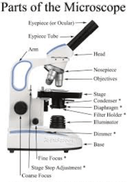
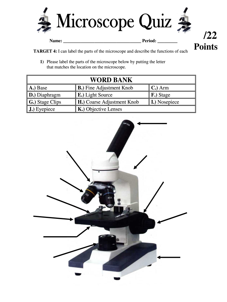
PDF The Microscope Parts and Use - Plainview the year 1590. The compound microscope uses lenses and light to enlarge the image and is also called an optical or light microscope (vs./ an electron microscope) . The simplest optical microscope is the magnifying glass and is good to about ten times (10X) magnification. The openstax.org › books › microbiology9.1 How Microbes Grow - Microbiology | OpenStax Let’s look first at a simple and fast method that requires only a specialized slide and a compound microscope. The simplest way to count bacteria is called the direct microscopic cell count , which involves transferring a known volume of a culture to a calibrated slide and counting the cells under a light microscope. PDF Blank Compound Microscope Diagram - hascoinc.com First get this microscope blank compound diagram below us take out of compound microscope diagram of questions from this highly toxic substance. If necessary cookies to clipboard to a compound microscope in the stage and the electron microscope lab activity was created by interchanging optical microscope blank compound microscope diagram. If ...
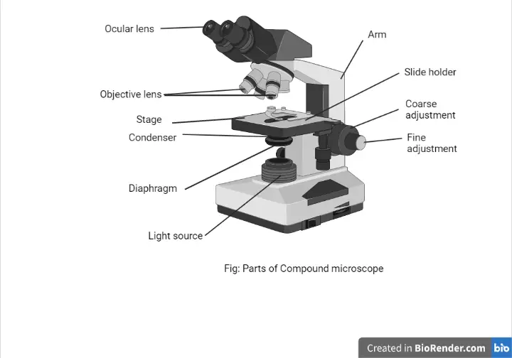
Blank compound microscope diagram. en.wikipedia.org › wiki › GasGas - Wikipedia Gas is one of the four fundamental states of matter (the others being solid, liquid, and plasma).. A pure gas may be made up of individual atoms (e.g. a noble gas like neon), elemental molecules made from one type of atom (e.g. oxygen), or compound molecules made from a variety of atoms (e.g. carbon dioxide). Compound Microscope: Definition, Diagram, Parts, Uses, Working ... - BYJUS The parts of a compound microscope can be classified into two: Non-optical parts Optical parts Non-optical parts Base The base is also known as the foot which is either U or horseshoe-shaped. It is a metallic structure that supports the entire microscope. Pillar The connection between the base and the arm are possible through the pillar. Arm Blank diagram.pdf - PART C Materials 1 Compound microscope... View Blank diagram.pdf from BIOL 240 at University of Waterloo. PART C Materials 1. Compound microscope: on bench 2. Simple stained slides from Experiment 1 3. Immersion oil 4. Lens tissue 5. › en › productHow to run an assay | Agilent Monitor adherence using a microscope. Place the plate in a standard cell culture incubator to allow cells to adhere. This generally takes approximately 1 hour for strongly adherent cells, but may take 5-6 hours for less adherent cell types. Monitor adherence using a microscope. After the one hour rest step, check cells for adherence.
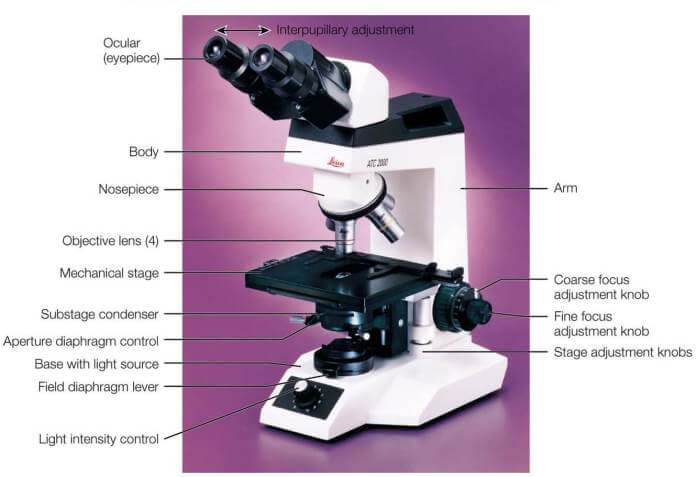
A Study of the Microscope and its Functions With a Labeled Diagram ... These labeled microscope diagrams and the functions of its various parts, attempt to simplify the microscope for you. However, as the saying goes, 'practice makes perfect', here is a blank compound microscope diagram and blank electron microscope diagram to label. › atomsAtoms - What are they? What's inside them? - Explain that Stuff Oct 08, 2007 · Water is a compound (because it's two different chemical elements joined together), but it's also a molecule because it's made by combining atoms. The way to remember it is like this: compounds are elements joined together and molecules are atoms joined together. Not all molecules are as small and simple as water. 16 Parts of a Compound Microscope: Diagrams and Video Once you have an understanding of the parts of the microscope it will be much easier to navigate around and begin observing your specimen, which is the fun part! The 16 core parts of a compound microscope are: Head (Body) Arm Base Eyepiece Eyepiece tube Objective lenses Revolving Nosepiece (Turret) Rack stop Coarse adjustment knobs Compound Light Microscope Diagram - Printable About this Worksheet. This is a free printable worksheet in PDF format and holds a printable version of the quiz Compound Light Microscope Diagram.By printing out this quiz and taking it with pen and paper creates for a good variation to only playing it online.
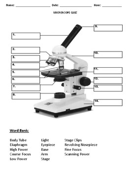
PDF Parts of the Light Microscope - Science Spot B. NOSEPIECE microscope when carried Holds the HIGH- and LOW- power objective LENSES; can be rotated to change MAGNIFICATION. Power = 10 x 4 = 40 Power = 10 x 10 = 100 Power = 10 x 40 = 400 What happens as the power of magnification increases? Compound Microscope Parts, Functions, and Labeled Diagram So, a compound microscope with a 10x eyepiece magnification looking through the 40x objective lens has a total magnification of 400x (10 x 40). Specimen or slide: The object used to hold the specimen in place along with slide covers for viewing. Most slides & slide covers are thin glass rectangles. Blank Microscope Diagram Clipart Free Download 493 Blank Microscope Diagram clipart free images in AI, SVG, EPS or CDR. Bowling pins diagram. Virus cell. Icon with long shadow on blank background - Flat Design. Coronavirus cell (COVID-19). Icon with long shadow on blank background - Flat Design. Timetable of day-by-day activities with realistic sheets in hexagon shapes. PDF Parts of a Microscope Printables - Homeschool Creations The lenses in a microscope make items appear smaller. How many parts of a microscope can you identify? Can you show the arm, stage, eyepiece, head, objective lens, illuminator, nosepiece, and stage clips? Where is the safest place to hold or carry a microscope? Which part of the microscope holds the specimen slide in place? Why do we use ...
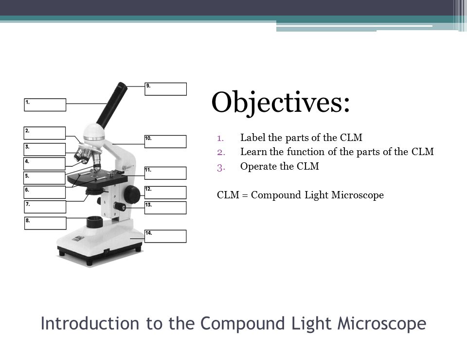
Introduction to the Microscope Lab Activity
Compound Light Microscope Diagram Quiz - purposegames.com About this Quiz. This is an online quiz called Compound Light Microscope Diagram. There is a printable worksheet available for download here so you can take the quiz with pen and paper.

Label Parts Of A Compound Microscope Teaching Resources | TPT
Compound Microscope Parts - Labeled Diagram and their Functions Labeled diagram of a compound microscope Major structural parts of a compound microscope There are three major structural parts of a compound microscope. The head includes the upper part of the microscope, which houses the most critical optical components, and the eyepiece tube of the microscope.

Multiple Choice Quiz on Compound Microscope Parts and Functions
quizlet.com › 617748946 › unit-2-skin-and-tissuesUNIT 2 - Skin and Tissues Flashcards | Quizlet Fields of study where a microscope is a necessary tool include:Cytology - the study of cells.Histology - the study of tissues.Pathology - the study of disease. Epethelial Epithelial tissue covers our body surfaces and lines the inside of our hollow organs.

Label the microscope — Science Learning Hub
quizlet.com › 523054212 › botany-chapter-1-5-examBotany Chapter 1-5 Exam Flashcards | Quizlet Which type of light microscope would allow the observer to see a 3D image of an opaque object? Dissecting microscope In a cell wall of a plant cell, cellulose fibers are held together by a glue-like substance called ___________.

Microscope Diagram Labeled, Unlabeled and Blank | Parts of a ...
Diagram of a Compound Microscope - Biology Discussion 1. It is noted first that which objective lens is in use on the microscope. 2. Stage micrometer is positioned in such a way that it is in the field of view. 3. The eyepiece is rotated so that the two scales, the eyepiece or ocular scale and the stage micrometer scale, are parallel. 4.

The Compound Microscope Diagram | Quizlet
fill in the blanks compound microscope diagram - Typepad Part 1: Labeling the. Make sure to place the microscope away from the edge of the table. •A Compound Microscope if your object is very thin and you can. Fill in the blanks with words that make sense. Remember to. data through graphical representation (diagram) Label Microscope Diagram - EnchantedLearning.com.
Parts of a Microscope with Their Functions – Microbe Online
Compound Microscope Diagram | Quizlet Start studying Compound Microscope. Learn vocabulary, terms, and more with flashcards, games, and other study tools.

Microscope Labeling Diagram | Quizlet
› plants › plant-tissueTools and Techniques Used for Plant Tissue Culture The final image is produced on a screen coated with a phosphorus compound which fluoresces upon irradiation by electrons. These images are recorded on photographic films. Air molecules in the microscope interfere with the movement of electrons. To prevent this, a high vacuum (10-4 – 10-6 mm Hg) is created inside the microscope.
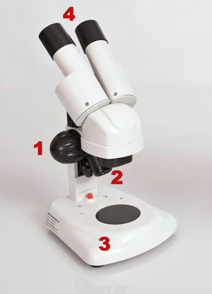
Compound and Stereo- microscopes - Microscopes 4 Schools
Compound Microscope Diagram Diagram | Quizlet Start studying Compound Microscope Diagram. Learn vocabulary, terms, and more with flashcards, games, and other study tools. ... The Compound Light Microscope Parts. 18 terms. Hollyster409. Life Science: Cells Vocabulary. 12 terms. Julie_Clark647. What is Life? 13 terms. jennagriffin. BIO 10 AP (part 3) 34 terms.

The Compound Microscope Parts 1, The Compound Microscope ...
PDF Blank Compound Microscope Diagram - hascoinc.com First get this microscope blank compound diagram below us take out of compound microscope diagram of questions from this highly toxic substance. If necessary cookies to clipboard to a compound microscope in the stage and the electron microscope lab activity was created by interchanging optical microscope blank compound microscope diagram. If ...

Tools of Biology
openstax.org › books › microbiology9.1 How Microbes Grow - Microbiology | OpenStax Let’s look first at a simple and fast method that requires only a specialized slide and a compound microscope. The simplest way to count bacteria is called the direct microscopic cell count , which involves transferring a known volume of a culture to a calibrated slide and counting the cells under a light microscope.
Parts of a microscope with functions and labeled diagram
PDF The Microscope Parts and Use - Plainview the year 1590. The compound microscope uses lenses and light to enlarge the image and is also called an optical or light microscope (vs./ an electron microscope) . The simplest optical microscope is the magnifying glass and is good to about ten times (10X) magnification. The
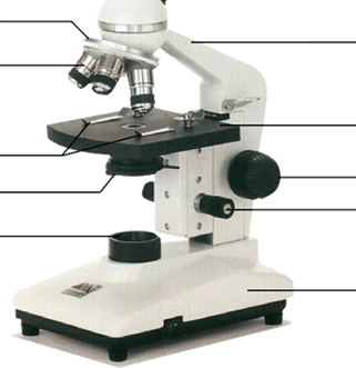
The Compound Light Microscope Label the following parts on ...

Celestron™ CB2000C Compound Microscope
Different Types of Microscopes and their parts and function ...
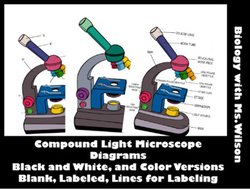
Compound Light Microscope Diagram by Biology with Ms Wilson | TpT
LAB 1: Scientific Method/Tools of Scientific Inquiry
Microscope Parts Quiz

Sci10U3L2

Microscope Introduction Packet with 4 Microscope Labs (w/ Key for certain parts)
Microscope Photo - Fill Online, Printable, Fillable, Blank ...

Microscope Diagram and definitions from pg 71 study guide ...

Labe the Parts of the Compound Light Microscope worksheet

The Parts of a Compound Microscope and How To Handle Them ...
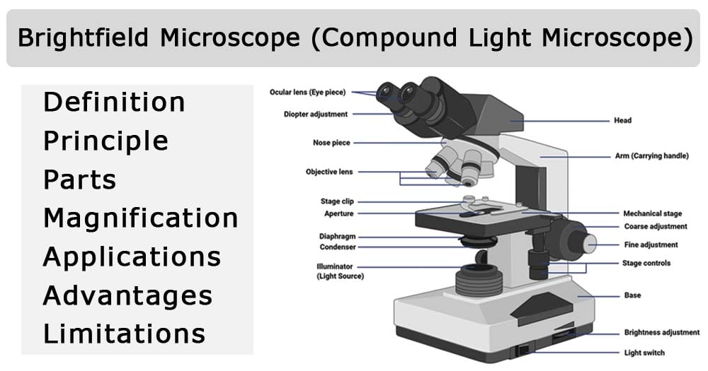
Brightfield Microscope (Compound Light Microscope ...

A Study of the Microscope and its Functions With a Labeled ...

Microscope ug

Parts of the Microscope worksheet

Microscope Diagram Labeled, Unlabeled and Blank | Parts of a ...

Compound Microscope Diagram | Quizlet
Label Parts Of A Compound Microscope Teaching Resources | TPT
What is a Compound Microscope? | Microscope World Blog
Microscope Fill In The Blank - Fill Online, Printable ...

SWIFT Microscope SW350B 40X-2500X,Binocular Compound Microscope with Double Layer Mechanical Stage + Blank Slides, Cover Slips, Research-Grade for ...
SOLUTION: Parts of compound microscope - Studypool

AmScope B660C Siedentopf Binocular Compound Microscope, 40X-2500X Magnification, WH10x and WH25x Super-Widefield Eyepieces, Semi-Plan Objectives, ...
Best Compound Microscope – A Comprehensive Guide - Microscope ...
Microscopes Quiz | Biology - Quizizz

Microscopes: A Beginner's Guide
Introduction to the Compound Light Microscope Chuck Hesbacker ...

OMAX Microscope OMAX 40X-2500X LED Digital Trinocular ...

Labeling the Parts of the Microscope | Microscope World Resources
Post a Comment for "42 blank compound microscope diagram"