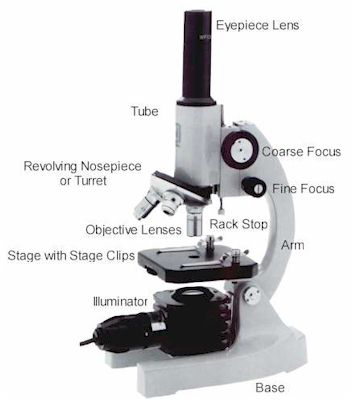
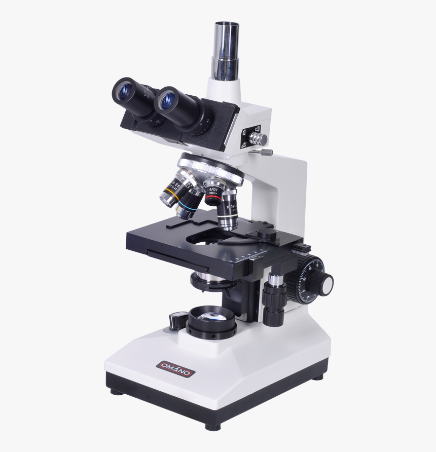
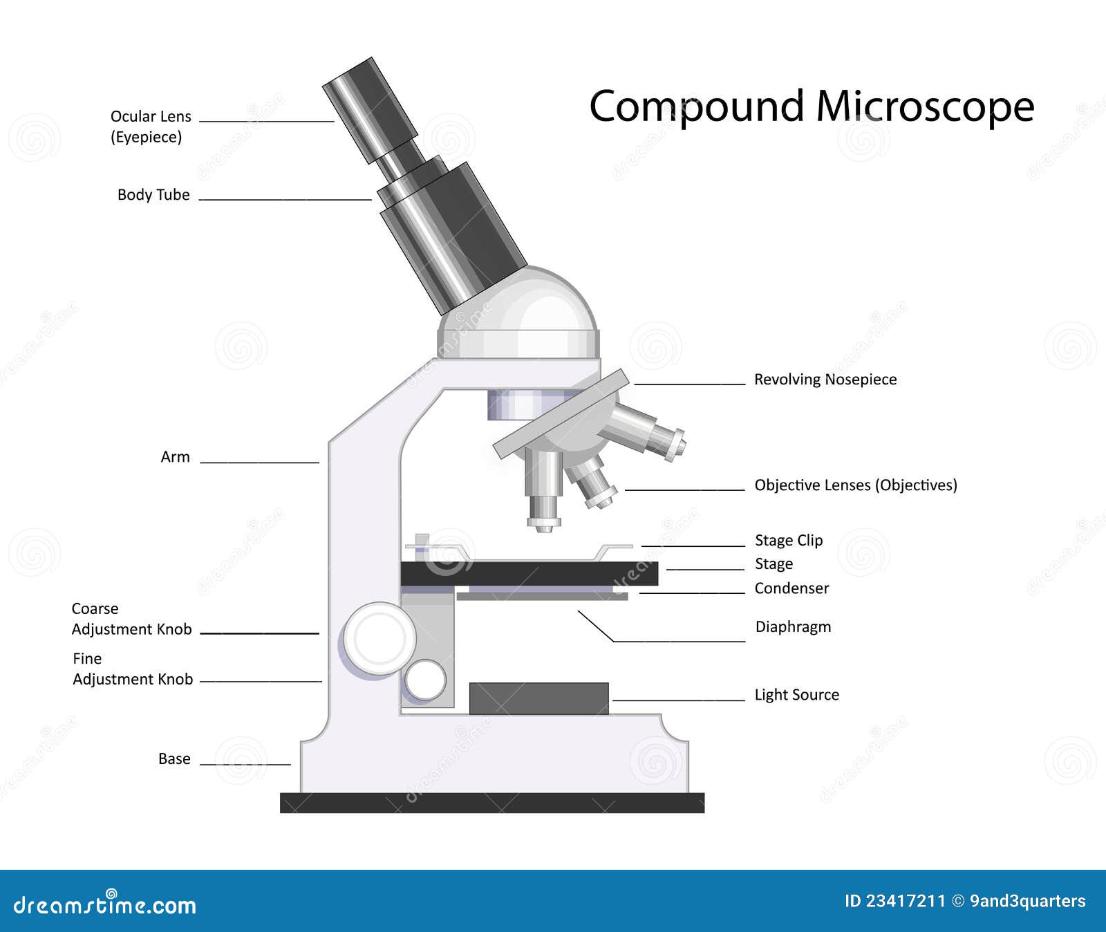
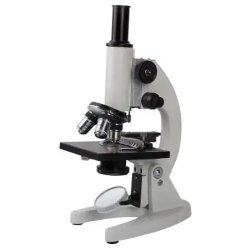
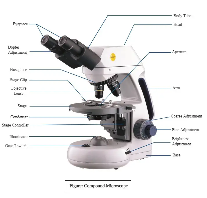
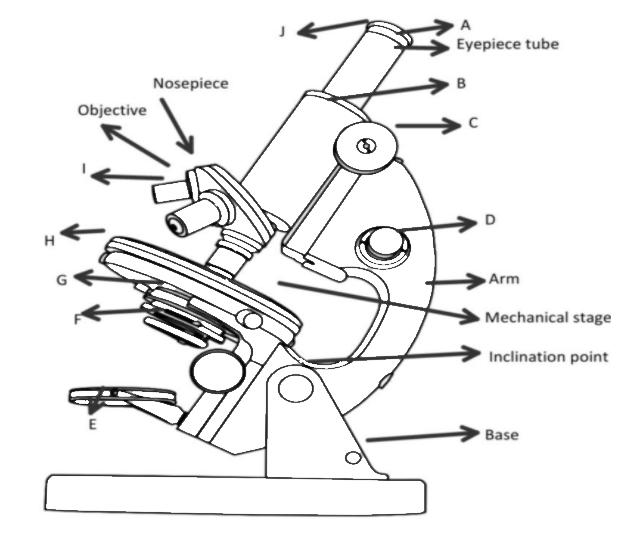
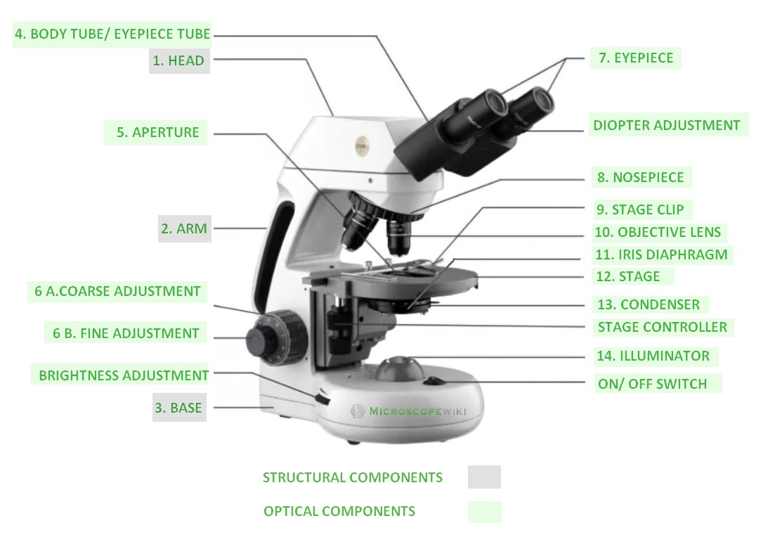
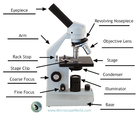
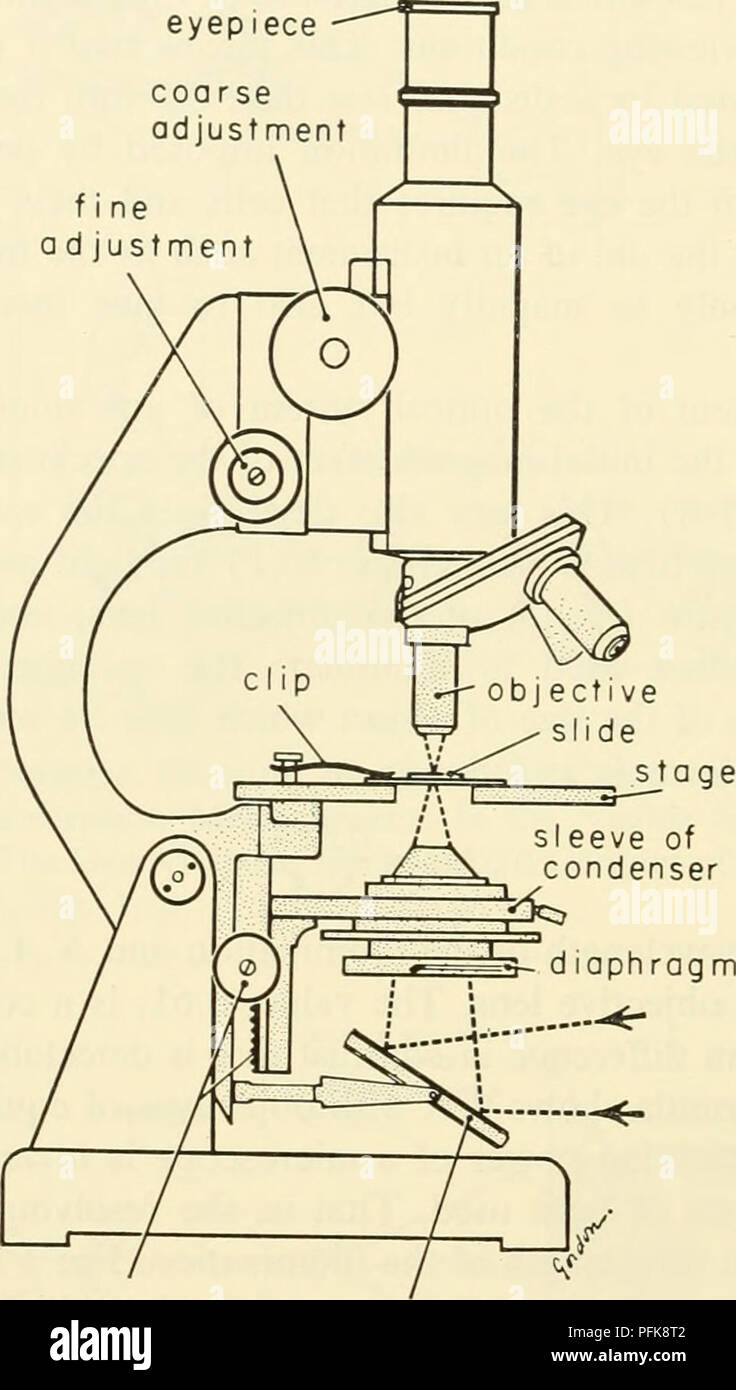
45 labeled compound microscope
› parts-of-a-compoundMicroscope Parts and Functions With Labeled Diagram and ... Before exploring microscope parts and functions, you should probably understand that the compound light microscope is more complicated than just a microscope with more than one lens. First, the purpose of a microscope is to magnify a small object or to magnify the fine details of a larger object in order to examine minute specimens that cannot ... Experiment 1 ( Cheah Kheng Keat H8T03B) PDF - Flip eBook Pages 1-9 ... Light compound microscope is a microscope that use light source and has 2 set of lenses, ocular lens and objective lens. We should always hold the microscope with both hands, that is hold the body arm of the microscope with one hand and the base of the microscope with the other hand. We use the oil immersion objective lens to observe
Light Microscope (Theory) - Amrita Vishwa Vidyapeetham Light microscope uses the properties of light to produce an enlarged image. It is the simplest type of microscope. Based on the simplicity of the microscope it may be categorized into: A) Simple microscope. B) Compound microscope. A) Simple microscope . It is uses only a single lens, e.g.: hand lens. Most of these are double convex or ...
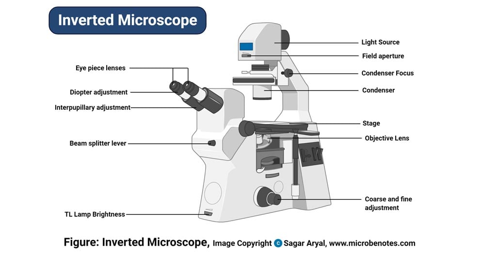
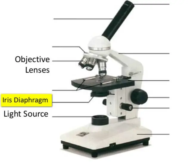
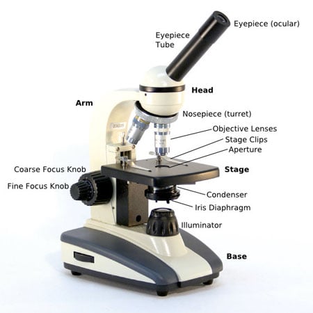
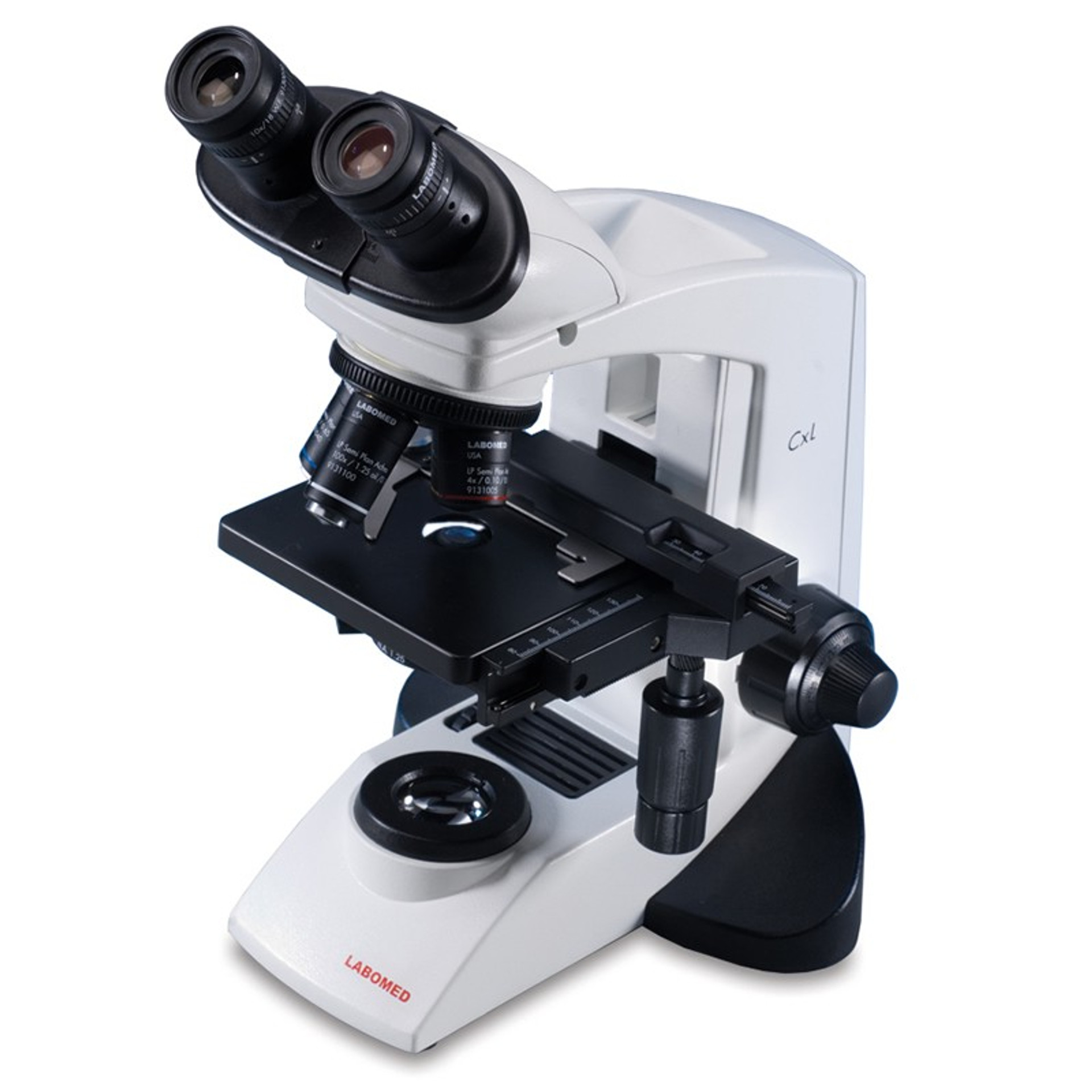
Labeled compound microscope
Welcome to microscopy4kids - Microscopy4kids Welcome to microscopy4kids! Your microscope guide and resource are here. Microscopy4kids are currently maintained by Rs Science. How to observe sugar under a microscope? Sugar and artificial sweetener. Sugar is a great subject for young kids and microscope enthusiasts to start their exploration. Different types of sugars and …. Parts of the Microscope with Labeling (also Free Printouts) Parts of the Microscope with Labeling (also Free Printouts) By Editorial Team March 7, 2022 A microscope is one of the invaluable tools in the laboratory setting. It is used to observe things that cannot be seen by the naked eye. Table of Contents 1. Eyepiece 2. Body tube/Head 3. Turret/Nose piece 4. Objective lenses 5. Knobs (fine and coarse) 6. Gen-Bio-1_Compound-Microscope-Activity-Gertos.docx - Name:... Name: ERL JOHN GERTOS _____ Date Performed: August 22, 2022 Date Submitted: August 22, 2022 Activity No. 1 THE COMPOUND MICROSCOPE The microscope is an essential tool in the study of the fine structure of animals. It enables you to see the animal structures too small to be seen with the naked eye. The microscope is made up of a system of lenses that can invert images.
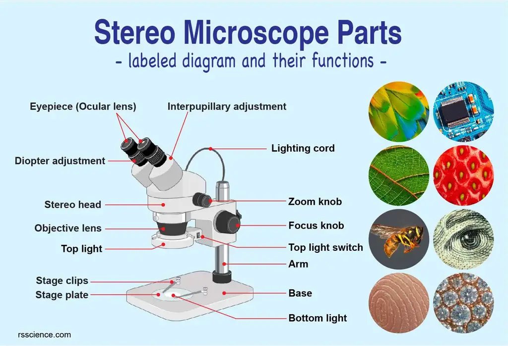
Labeled compound microscope. Microscopy- History, Classification, Terms, Diagram - The Biology Notes A simple or compound light microscope is used in this technique. It uses transmitted visible light to develop magnified images. It has a low contrasting capacity, low optical resolution, requires staining and has a limited magnification of around 1300X. It is simple and can be used to observe living cells and microorganisms. 2 Dark Field Microscopy rsscience.com › stereo-microscopeParts of Stereo Microscope (Dissecting microscope) – labeled ... If you would like to learn optical components of a compound microscope, please visit Compound Microscope Parts – Labeled Diagram and their Functions, and this article. How to use a stereo (dissecting) microscope. Follow these steps to put your stereo microscopes in work: 1. Set your microscope on a tabletop or other flat sturdy surface where ... Parts of Stereo Microscope (Dissecting microscope) – labeled … If you would like to learn optical components of a compound microscope, please visit Compound Microscope Parts – Labeled Diagram and their Functions, and this article. How to use a stereo (dissecting) microscope. Follow these steps to put your stereo microscopes in work: 1. Set your microscope on a tabletop or other flat sturdy surface where ... UD Virtual Compound Microscope - University of Delaware ©University of Delaware. This work is licensed under a Creative Commons Attribution-NonCommercial-NoDerivs 2.5 License.Creative Commons Attribution-NonCommercial-NoDerivs 2.5 …
A Guide to Different Microscope Types and Their Various Uses A compound microscope might also be labeled as a biological microscope. Compound microscopes are used in laboratories, wastewater treatment plants, schools, and more. Samples viewed under a compound microscope have to be prepared on a microscope slide using a coverslip to help flatten the sample. CBSE Class 9 Science Practicals Identification of parenchyma ... 2) Adjust the slide under compound microscope. 3) Initially adjust the slide on 10 X objective and then 45X objective, 4) Draw the diagram of your observation and label appropriately. Observation: a) For identification of parenchyma following characteristics were observed. 1) Presence of intra cellular spaces in parenchymal cells. microbenotes.com › compound-microscope-principleCompound Microscope- Definition, Labeled Diagram, Principle ... Apr 03, 2022 · Working Principle of the Compound Microscope Compound microscopes have a combination of lenses that enhances both magnifying powers as well as the resolving power. The specimen or object, to be examined is usually mounted on a transparent glass slide and positioned on the specimen stage between the condenser lens and objective lens. How does a Microscope work With a compound microscope, the total magnification can be determined by multiplying the magnifications of the objective and ocular lenses. Consequently, an ocular lens of 10X coupled with a 40X objective yields a total magnification of 400X. However, the higher the magnification the closer the lens must be to the specimen. Since a higher magnification lens bends light …
researchtweet.com › microscope-parts-labeledMicroscope, Microscope Parts, Labeled Diagram, and Functions Jan 19, 2022 · Simply multiply the magnification of the ocular lens by the magnification of the objective lens to calculate the power of magnification of a microscope. For a typical compound microscope with a 10X ocular lens and objective lenses with magnifications of 4X, 10X, 40X, and 100X, your microscope will have magnifications of 40X, 100X, 400X, and ... Metabolic labelling of cancer cells with glycodendrimers stimulate ... Strategy combining glycometabolism and bio-orthogonal click chemistry to label cells with clustered rhamnose antigen and activate immune response against cancer cells. ... (20.0 mg, 2.2 μmol). The crude mixture was purified (0-80% B in 20 min) to afford the title compound (15.1 mg, 1.5 μmol, 71%). ... Spheroids were imaged using confocal ... Safety of use of the ENDOSWIR near-infrared optical ... - SpringerLink The evaluation was made on Formalin-Fixed Paraffin-Embedded (FFPE) samples using an Olympus optical microscope at × 400 magnification, field diameter 0.62 mm, after Hematoxylin Eosin Safran (HES) staining. ... J. Olson, and F. Robles, (2021) Label-free detection of brain tumors in a 9L gliosarcoma rat model using stimulated Raman scattering ... An Introduction to the Light Microscope, Light Microscopy Techniques ... Therefore, the total magnification of a compound microscope, which is the product of the objective magnification and the eyepiece magnification, typically lies in the range of 400-1000 x. ... Examples include biological samples that are intrinsically fluorescent or have been labeled with a fluorescent marker, as well as single molecules and ...
Microscope Types (with labeled diagrams) and Functions Compound microscope labeled diagram Compound microscope functions: It finds great application in areas of pathology, pedology, forensics etc Its greater order of magnification allows for deeper study of microbial organisms to Detect the cause of diseases Study the mineral composition in soils
Compound Microscope- Definition, Labeled Diagram, Principle, … 03/04/2022 · Magnification of compound microscope In order to ascertain the total magnification when viewing an image with a compound light microscope, take the power of the objective lens which is at 4x, 10x or 40x and multiply it by the power of the eyepiece which is typically 10x.
Microscope: Types of Microscope, Parts, Uses, Diagram - Embibe Compound Microscope. A compound microscope is defined as a microscope with a high resolution. It uses two sets of lenses, providing a \(2\)-dimensional image of the sample. The term compound refers to the usage of more than one lens in the microscope. Also, the compound microscope is one of the types of optical microscopes.
Microscope Parts, Function, & Labeled Diagram - slidingmotion Microscope parts labeled diagram gives us all the information about its parts and their position in the microscope. Microscope Parts Labeled Diagram The principle of the Microscope gives you an exact reason to use it. It works on the 3 principles. Magnification Resolving Power Numerical Aperture. Parts of Microscope Head Base Arm Eyepiece Lens
Diatoms Under A Microscope Labeled - chunyinga.blogspot.com Diatoms Under A Microscope Labeled Displaying Images For Metabolic Engineering Of Tio2 Nanoparticles In Nitzschia Palea To Terrestrial Diatoms As. Elegans was genetically labeled with green fluorescent proteins GFP in all the cells. Valves with radial symmetry symmetric about a point Cells lack a raphe system and lack significant motility.
Microscope, Microscope Parts, Labeled Diagram, and Functions 19/01/2022 · For a typical compound microscope with a 10X ocular lens and objective lenses with magnifications of 4X, 10X, 40X, and 100X, your microscope will have magnifications of 40X, 100X, 400X, and 1000X depending on which objective lens you use. The same principle applies to stereo microscopes; a 10X eyepiece combined with a 4X objective lens produces a …
Parts of a microscope with functions and labeled diagram 19/04/2022 · Q. Differentiate between a condenser and an Abbe condenser. Ans. Condensers are lenses that are used to collect and focus light from the illuminator into the specimen. They are found under the stage next to the diaphragm of the microscope. They play a major role in ensuring clear sharp images are produced with a high magnification of 400X and above.
Compound Microscope - Types, Parts, Diagram, Functions and Uses Compound microscope - It is an optical instrument consists of two convex lenses of short focal lengths primarily used for observing a highly magnified image of minute objects. Lenses Simple microscope - It has a convex lens. It uses only one lens to magnify objects. An example of a simple microscope is a magnifying glass.
Microscopic Morphology - BIO 2410: Microbiology - Baker College Eukaryotic microbes include protists, some fungi, and some flatworms (platyhelmintes) and roundworms (nematodes). The powerpoint file below introduces you to some of these Eukaryotic microbes. After reviewing the powerpoint file, see if you can identify the eukaryotic microbes in the fungi, protists, and helminth categories.
What is a Stereo Microscope? - New York Microscope Company 11/05/2018 · Each part of a stereo microscope is labeled in the diagram below. This example is a typical classroom type stereo microscope with track stand and built-in illumination. Stereo Head: This is the moveable top portion of the microscope and the stereo head holds the two adjustable eyepieces. Ocular Lens: These are the eyepieces through which the viewer looks at the …
Microscope Parts and Functions Microscope Parts and Functions With Labeled Diagram and Functions How does a Compound Microscope Work?. Before exploring microscope parts and functions, you should probably understand that the compound light microscope is more complicated than just a microscope with more than one lens.. First, the purpose of a microscope is to magnify a …
The Compound Microscope - Apps on Google Play $2.49 Buy About this app arrow_forward "Compound Microscope" app brings to you a guided tour to acquaint yourself about Compound Microscope. The app brings to your finger tip the step by step...
MCQ on Microscopy Pdf - YB Study The image formed by an objective of a compound microscope is real and enlarged. The electron microscope is the most powerful. The principle of an electron microscope is the same as that of an optical microscope, except that the electrons have shorter wavelengths and can see smaller objects.
Microscopy Pre-lab Activities - University of Delaware Microscope controls: turn knobs (click and hold on upper or lower portion of knob) throw switches (click and drag) turn dials (click and drag) move levers (click and drag) changes lenses (click and drag on objective housing) select a specimen (click on a slide) adjust oculars (in "through" view, with the light on, click and drag to move oculars closer or further apart) Designed and …
Microscope Quiz: How Much You Know About Microscope Parts ... - ProProfs Projects light upwards through the diaphragm, the specimen, and the lenses. 5. Is used to regulates the amount of light on the specimen. Supports the slide being viewed. Moves the stage up and down for focusing. 6. Is used to support the microscope when carried. Moves the stage slightly to sharpen the image.
microscopewiki.com › compound-microscopeCompound Microscope – Diagram (Parts labelled), Principle and ... Also called as binocular microscope or compound light microscope, it is a remarkable magnification tool that employs a combination of lenses to magnify the image of a sample that is not visible to the naked eye. Compound microscopes find most use in cases where the magnification required is of the higher order (40 - 1000x).
Compound Microscope – Diagram (Parts labelled), Principle and … 03/02/2022 · Image : Labeled Diagram of compound microscope parts. See: Labeled Diagram showing differences between compound and simple microscope parts Structural Components. The three structural components include. 1. Head. This is the upper part of the microscope that houses the optical parts. 2. Arm . This part connects the head with the base and provides …
microbenotes.com › parts-of-a-microscopeParts of a microscope with functions and labeled diagram Apr 19, 2022 · Figure: Diagram of parts of a microscope. There are three structural parts of the microscope i.e. head, base, and arm. Head – This is also known as the body. It carries the optical parts in the upper part of the microscope. Base – It acts as microscopes support. It also carries microscopic illuminators.
www1.udel.edu › biology › ketchamUD Virtual Compound Microscope - University of Delaware ©University of Delaware. This work is licensed under a Creative Commons Attribution-NonCommercial-NoDerivs 2.5 License.Creative Commons Attribution-NonCommercial-NoDerivs 2
39 Fun Facts about Light, Compound and Electron Microscopes Carl Zeiss binocular compound microscope, 1914 Fun facts about microscopes 1. Cornelis Drebbel shows a compound microscope with a convex objective and a convex eyepiece in London in 1621. (a "Keplerian" microscope). Drebbel exhibits his invention in Rome in c.1622. 2.
Substrate-bound and substrate-free outward-facing structures of a ... Crystal structure of BmrA in OF conformation in complex with ATP-Mg 2+. We first stabilized BmrA in its OF conformation by introducing the E504A mutation that prevents hydrolysis of ATP (25, 31) and doxorubicin transport (fig. S1).The protein crystallized following the procedure setup for the mouse P-glycoprotein, using Triton X-100 for extraction and a mixture of N-dodecyl-β-d-n ...
Amoeba Under Microscope 40X Labeled - Amoeba Cell / This is a great ... Add a labeled scale bar to the drawing of the same amoeba, then determine the . Scanning (4x), low (10x), high (40x), and oil immersion (100x). Your microscope has 4 objective lenses: To drop of water on slide, mix, cover w/ coverslip, observe under compound microscope. Calculate the field of view diameter of a microscope under medium or high.
Gen-Bio-1_Compound-Microscope-Activity-Gertos.docx - Name:... Name: ERL JOHN GERTOS _____ Date Performed: August 22, 2022 Date Submitted: August 22, 2022 Activity No. 1 THE COMPOUND MICROSCOPE The microscope is an essential tool in the study of the fine structure of animals. It enables you to see the animal structures too small to be seen with the naked eye. The microscope is made up of a system of lenses that can invert images.
Parts of the Microscope with Labeling (also Free Printouts) Parts of the Microscope with Labeling (also Free Printouts) By Editorial Team March 7, 2022 A microscope is one of the invaluable tools in the laboratory setting. It is used to observe things that cannot be seen by the naked eye. Table of Contents 1. Eyepiece 2. Body tube/Head 3. Turret/Nose piece 4. Objective lenses 5. Knobs (fine and coarse) 6.
Welcome to microscopy4kids - Microscopy4kids Welcome to microscopy4kids! Your microscope guide and resource are here. Microscopy4kids are currently maintained by Rs Science. How to observe sugar under a microscope? Sugar and artificial sweetener. Sugar is a great subject for young kids and microscope enthusiasts to start their exploration. Different types of sugars and ….













Post a Comment for "45 labeled compound microscope"