42 microscope diagram labelled
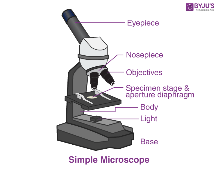
Investigating cells with a light microscope - BBC Bitesize Record the microscope images using labelled diagrams or produce digital images. When first examining cells or tissues with low power, draw an image at this stage, even if … Microscope Types (with labeled diagrams) and Functions Simple microscope labeled diagram Simple microscope functions It is used in industrial applications like: Watchmakers to assemble watches Cloth industry to count the number of threads or fibers in a cloth Jewelers to examine the finer parts of jewelry Miniature artists to examine and build their work Also used to inspect finer details on products
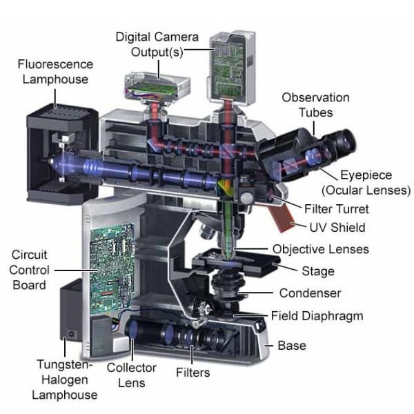
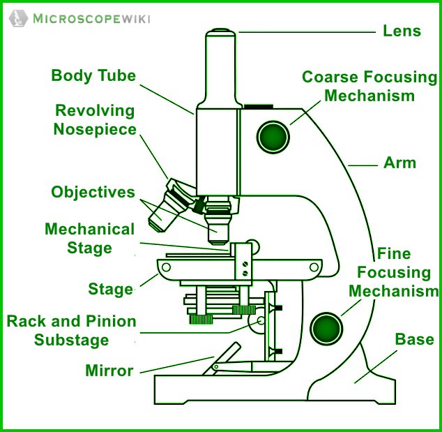
Compound Microscope Parts - Labeled Diagram and their Functions Labeled diagram of a compound microscope Major structural parts of a compound microscope There are three major structural parts of a compound microscope. The head includes the upper part of the microscope, which houses the most critical optical components, and the eyepiece tube of the microscope.

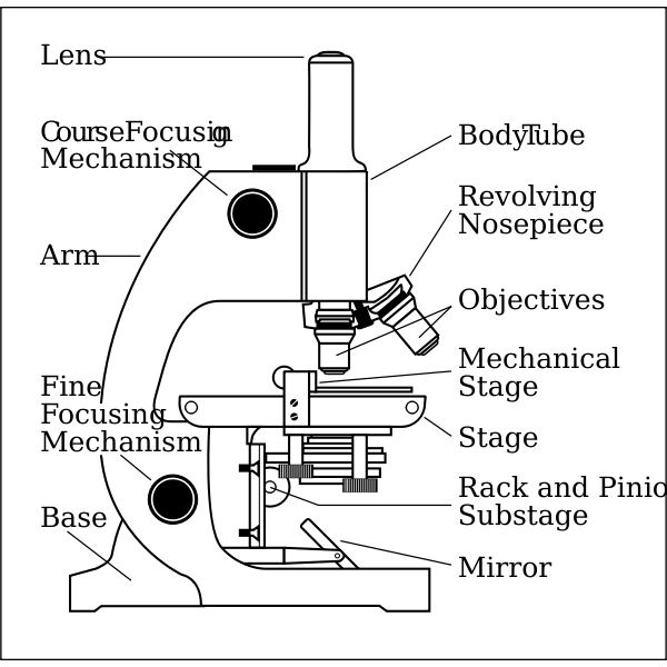
Microscope diagram labelled
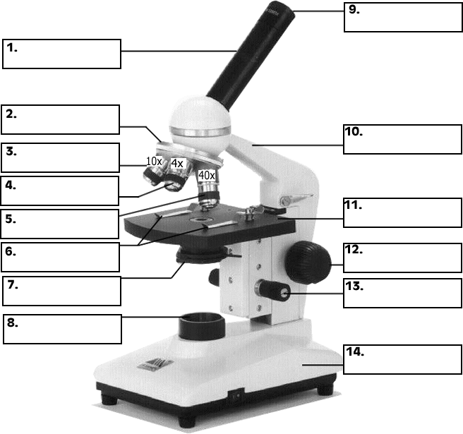
Labeling the Parts of the Microscope | Microscope World Resources Labeling the Parts of the Microscope This activity has been designed for use in homes and schools. Each microscope layout (both blank and the version with answers) are available as PDF downloads. You can view a more in-depth review of each part of the microscope here. Download the Label the Parts of the Microscope PDF printable version here. Required practical - using a light microscope - BBC Bitesize Record the microscope images using labelled diagrams or produce digital images. When first examining cells or tissues with low power, draw an image at this stage, even if … 4 Major Phases of the Cell Cycle (With Diagram) ADVERTISEMENTS: The following points highlight the four major phases of the cell cycle. The phases are: 1. G1 (gap1) phase 2. S (synthesis) phase 3. G2 (gap 2) phase 4. M (mitosis) phase. Cell Cycle: Phase # 1. G1 Phase: The G1 phase is set in immediately after the cell division. It is characterised by […]
Microscope diagram labelled. Core practical 3: Observe mitosis in root tips - Edexcel 5. After 5 minutes, use forceps to take the tips out of the vial, and place them on a microscope slide. Add a drop of water to the root tip on the slide. Tease the root tip apart with needles (maceration), to spread out the cells a little. Cover with a coverslip. Replace the lid on the vial of stain and return it to the teacher as instructed. 6 ... What is skin? The layers of human skin - YouTube The skin is the largest organ of the human body, weighing approximately 16% of our bodyweight. Skin consists of multiple layers, epidermis, dermis and hypode... Label Microscope Diagram - EnchantedLearning.com base - this supports the microscope. body tube - the tube that supports the eyepiece. coarse focus adjustment - a knob that makes large adjustments to the focus. diaphragm - an adjustable opening under the stage, allowing different amounts of light onto the stage. eyepiece - where you place your eye. Microscope, Microscope Parts, Labeled Diagram, and Functions Microscope, Microscope Parts, Labeled Diagram, and Functions What is Microscope? A microscope is a laboratory instrument used to examine objects that are too small to be seen by the naked eye. It is derived from Ancient Greek words and composed of mikrós, "small" and skopeîn,"to look" or "see".
Sperm Under Microscope with Labeled Diagram - AnatomyLearner The labelled diagram has already described all the structures of sperm in this article. Conclusion. So, this article provides the details structural features of sperm under the light microscope. All the labeled diagrams might help you identify the sperms from seminiferous tubules and epididymis of an animal. A Study of the Microscope and its Functions With a Labeled Diagram ... A Study of the Microscope and its Functions With a Labeled Diagram To better understand the structure and function of a microscope, we need to take a look at the labeled microscope diagrams of the compound and electron microscope. These diagrams clearly explain the functioning of the microscopes along with their respective parts. Electron microscope - Wikipedia An electron microscope is a microscope that uses a beam of accelerated electrons as a source of illumination. As the wavelength of an electron can be up to 100,000 times shorter than that of visible light photons, electron microscopes have a higher resolving power than light microscopes and can reveal the structure of smaller objects. Label the Microscope Diagram | Download Scientific Diagram - ResearchGate Label the Microscope Diagram Source publication +5 Laboratory Exercises in Microbiology: Discovering the Unseen World through Hands-on Investigation Book Full-text available Oct 2016 Joan Petersen...
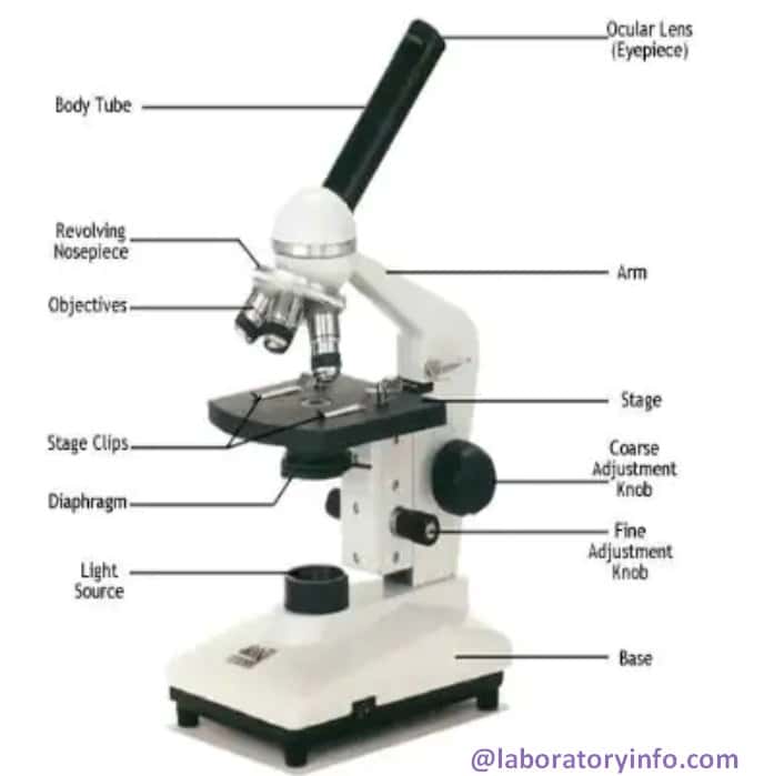
Microscope Diagram Labeled, Unlabeled and Blank | Parts of a Microscope ... Description Worksheet identifying the parts of the compound light microscope. Answer key: 1. Body tube 2. Revolving nosepiece 3. Low power objective 4. Medium power objective 5. High power objective 6. Stage clips 7. Diaphragm 8. Light source 9. Eyepiece 10. Arm 11. Stage 12. Coarse adjustment knob 13. Fine adjustment knob 14. Base S Top 16 Techniques Used in Cell Biology (With Diagram) Then the bound labelled antigens are eluted from the column and can be quantitatively arranged through Liquid Scintillation counter. Antibodies are generally labelled with H 3,C 14 or I 131. When cells or tissues axe used, radiolabeled antibodies can be used with the help of autoradiography to localize the various components within the cell. Compound Microscope- Definition, Labeled Diagram, Principle, Parts, Uses The naked eye can now view the specimen at magnification 400 times greater and so microscopic details are revealed. Alternatively, the magnification of the compound microscope is given by: m = D/ fo * L/fe where, D = Least distance of distinct vision (25 cm) L = Length of the microscope tube fo = Focal length of the objective lens Electron microscope - Wikipedia Diagram illustrating the phenomena resulting from the interaction of highly energetic electrons with matter. In 1926, Hans Busch developed the electromagnetic lens. According to Dennis Gabor, the physicist Leó Szilárd tried in 1928 to convince him to build an electron microscope, for which he had filed a patent. The first prototype electron microscope, capable of four …
Microscope Diagram Labeled, Unlabeled and Blank - Pinterest timvandevall.com Microscope Diagram Labeled, Unlabeled and Blank | Parts of a Microscope - Tim's Printables Print a microscope diagram, microscope worksheet, or practice microscope quiz in order to learn all the parts of a microscope. T Tim's Printables 37k followers More information Microscope Diagram
The Eyes (Human Anatomy): Diagram, Optic Nerve, Iris, Cornea ... - WebMD WebMD's Eyes Anatomy Pages provide a detailed picture and definition of the human eyes. Learn about their function and problems that can affect the eyes.
Label the microscope — Science Learning Hub Label the microscope Interactive Add to collection Use this interactive to identify and label the main parts of a microscope. Drag and drop the text labels onto the microscope diagram. high-power objective coarse focus adjustment stage base fine focus adjustment light source diaphragm or iris eye piece lens Download Exercise Tweet
Microscope Labeled Pictures, Images and Stock Photos photosynthesis. Diagram of the process of photosynthesis, showing the light reactions and the Calvin cycle. photosynthesis by absorbing water, light from the sun, and carbon dioxide from the atmosphere and converting it to sugars and oxygen. Light reactions occur in the thylakoid. Calvin Cycle occurs in the stoma. Neutrophil vector illustration.
Parts of the Microscope Label and Definition Diagram | Quizlet Start studying Parts of the Microscope Label and Definition. Learn vocabulary, terms, and more with flashcards, games, and other study tools.
Labeled Microscope and Basics of Life Diagram | Quizlet A microscope is an instrument widely to magnify and resolve the image of an object that is otherwise invisible to naked eye. For resolving the details of objects, which otherwise cannot be achieved by naked eye, a microscope is used. This set of flash cards will help the student to identify the different parts and function of the microscope.
Simple Microscope - Diagram (Parts labelled), Principle, Formula and Uses A simple microscope consists of Optical parts Mechanical parts Labeled Diagram of simple microscope parts Optical parts The optical parts of a simple microscope include Lens Mirror Eyepiece Lens A simple microscope uses biconvex lens to magnify the image of a specimen under focus.
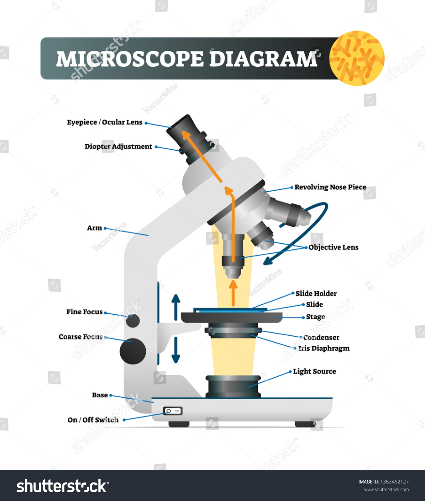
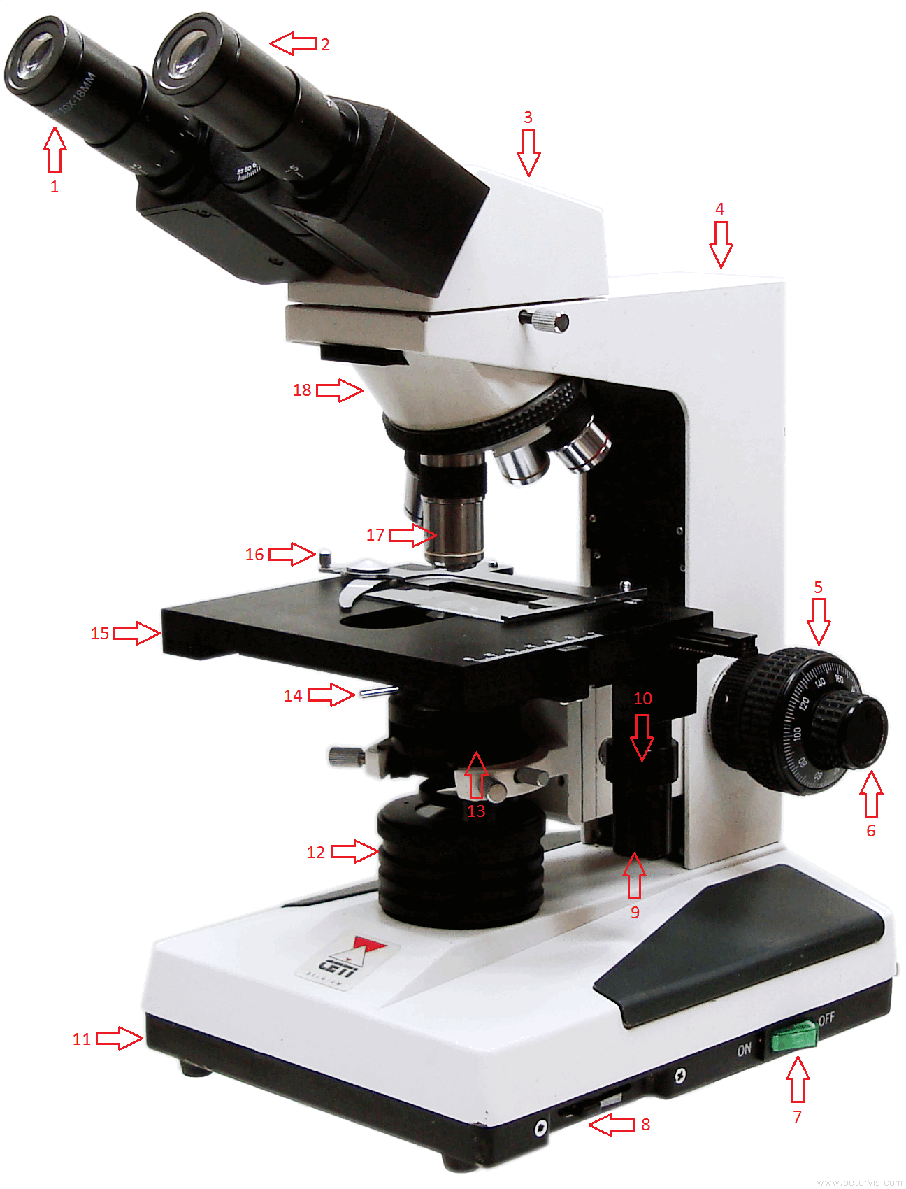
Parts of a microscope with functions and labeled diagram - Microbe Notes Figure: Diagram of parts of a microscope There are three structural parts of the microscope i.e. head, base, and arm. Head - This is also known as the body. It carries the optical parts in the upper part of the microscope. Base - It acts as microscopes support. It also carries microscopic illuminators.
Microscope Diagram Teaching Resources | Teachers Pay Teachers Teaching to the Middle. 4.8. (24) $1.50. PDF. This passage briefly describes microscopes and their parts (900-1000 Lexile). 14 questions (matching and multiple choice) assess students' understanding. Students label a diagram of 6 parts of a microscope. I've included a color and BW version, as well as a key.
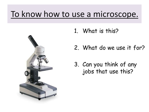
Microscope labeled diagram - slideshare.net Microscope labeled diagram 1. The Microscope Image courtesy of: Microscopehelp.com Basic rules to using the microscope 1. You should always carry a microscope with two hands, one on the arm and the other under the base. 2. You should always start on the lowest power objective lens and should always leave the microscope on the low power lens ...
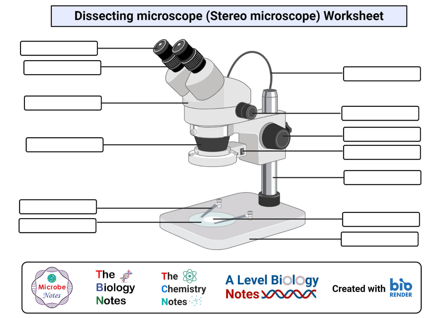
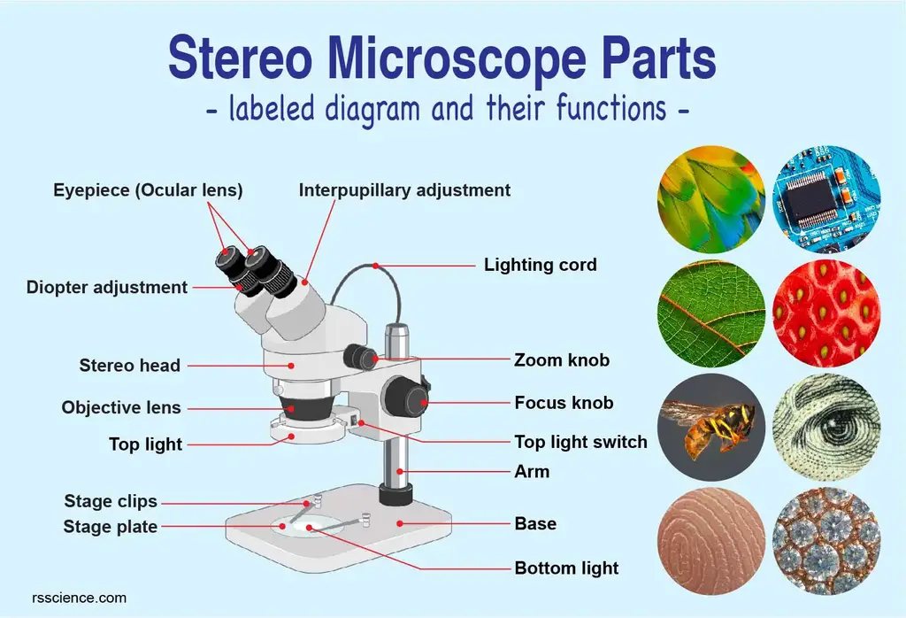
Parts of Stereo Microscope (Dissecting microscope) - labeled diagram ... Labeled part diagram of a stereo microscope Major structural parts of a stereo microscope There are three major structural parts of a stereo microscope. The viewing Head includes the upper part of the microscope, which houses the most critical optical components, including the eyepiece, objective lens, and light source of the microscope.
Microscope Parts, Function, & Labeled Diagram - slidingmotion Microscope parts labeled diagram gives us all the information about its parts and their position in the microscope. Microscope Parts Labeled Diagram The principle of the Microscope gives you an exact reason to use it. It works on the 3 principles. Magnification Resolving Power Numerical Aperture. Parts of Microscope Head Base Arm Eyepiece Lens
Neuron under Microscope with Labeled Diagram - AnatomyLearner Neuron under microscope labelled diagram. Throughout this article, you got the different neurons labelled diagrams. Here, you will also find the diagrams of different neuron types under a microscope. The neuron diagram shows the different parts (axon, dendrites, and cell body) of the neurons.
4 Major Phases of the Cell Cycle (With Diagram) - Biology … The following points highlight the four major phases of the cell cycle. The phases are: 1. G 1 (gap1) phase 2. S (synthesis) phase 3. G 2 (gap 2) phase 4. M (mitosis) phase. Cell Cycle: Phase # 1. G 1 Phase: . The G 1 phase is set in immediately after the cell division. It is characterised by a change in the chromosome from the condensed mitotic state to the more extended interphase …
pE-300white | LED Microscope Illuminator - CoolLED CoolLED Ltd supply LED Microscope Illuminator such as pE-300white for distributors throughout the world. Call +44 (0)1264 323040(UK) or 1.800.877.0128(USA).
Required practical - using a light microscope - Cells in ... Record the microscope images using labelled diagrams or produce digital images. When first examining cells or tissues with low power, draw an image at this stage, even if going on to examine the ...
PDF Label parts of the Microscope: Answers Label parts of the Microscope: Answers Coarse Focus Fine Focus Eyepiece Arm Rack Stop Stage Clip . Created Date: 20150715115425Z ...
Microscope Parts and Functions It also allows the specimen to be labeled, transported, and stored without damage. Stage: The flat platform where the slide is placed. Stage clips: Metal clips that hold the slide in place. Stage height adjustment (Stage Control): These knobs move the stage left and right or up and down.
Microscope Labeling - The Biology Corner Microscope Labeling. This simple worksheet pairs with a lesson on the light microscope, where beginning biology students learn the parts of the light microscope and the steps needed to focus a slide under high power. The labeling worksheet could be used as a quiz or as part of direct instruction where students label the microscope as you go ...
Investigating cells with a light microscope - BBC Bitesize Record the microscope images using labelled diagrams or produce digital images. When first examining cells or tissues with low power, draw an image at this stage, even if going on to examine the ...
Parts of a Compound Microscope and Their Functions - NotesHippo Compound microscope mechanical parts (Microscope Diagram: 2) include base or foot, pillar, arm, inclination joint, stage, clips, diaphragm, body tube, nose piece, coarse adjustment knob and fine adjustment knob.. Base: It's the horseshoe-shaped base structure of microscope.All of the other components of the compound microscope are supported by it. ...
Amazing 27 Things Under The Microscope With Diagrams 1. Amoeba under the microscope. Amoeba is a unicellular organism in the Kingdom Protozoa. It is a eukaryote and thus has membrane-bound cell organelles and protein-bound genetic material with a nuclear membrane. Amoeba moves with their pseudopodia, which is a specialized form of the plasma membrane that results in a crawling motion of the organism.
Top 16 Techniques Used in Cell Biology (With Diagram) ADVERTISEMENTS: The following points highlight the top sixteen techniques used in cell biology. Some of the techniques are: 1. Immunofluorescence Microscopy 2. Ion-Exchange Chromatography 3. Affinity Chromatography 4. Partition and Adsorption Chromatography 5. Gel Filtration Chromatography 6. Radioactive Tracer Technique 7. Radioimmunoassay (RIA) 8. …
Labelled Diagram of Compound Microscope - Biology Discussion The below mentioned article provides a labelled diagram of compound microscope. Part # 1. The Stand: The stand is made up of a heavy foot which carries a curved inclinable limb or arm bearing the body tube. The foot is generally horse shoe-shaped structure (Fig. 2) which rests on table top or any other surface on which the microscope in kept.
Compound Microscope Parts, Functions, and Labeled Diagram Compound Microscope Definitions for Labels Eyepiece (ocular lens) with or without Pointer: The part that is looked through at the top of the compound microscope. Eyepieces typically have a magnification between 5x & 30x. Monocular or Binocular Head: Structural support that holds & connects the eyepieces to the objective lenses.
Amazing 27 Things Under The Microscope With Diagrams 13/05/2022 · 1. Amoeba under the microscope. Amoeba is a unicellular organism in the Kingdom Protozoa. It is a eukaryote and thus has membrane-bound cell organelles and protein-bound genetic material with a nuclear membrane. Amoeba moves with their pseudopodia, which is a specialized form of the plasma membrane that results in a crawling motion of the organism.
22 Parts Of a Microscope With Their Function And Labeled Diagram The field diaphragm control is located around the lens located in the base. Hinge Screw -This screw fixes the arm to the base and allow for the tilting of the arm. Stage Clips - They hold the slide firmly onto the stage. On/OFF Switch - This switch on the base of the microscope turns the illuminator off and on.
What is skin? The layers of human skin - YouTube The skin is the largest organ of the human body, weighing approximately 16% of our bodyweight. Skin consists of multiple layers, epidermis, dermis and hypode...
4 Major Phases of the Cell Cycle (With Diagram) ADVERTISEMENTS: The following points highlight the four major phases of the cell cycle. The phases are: 1. G1 (gap1) phase 2. S (synthesis) phase 3. G2 (gap 2) phase 4. M (mitosis) phase. Cell Cycle: Phase # 1. G1 Phase: The G1 phase is set in immediately after the cell division. It is characterised by […]
Required practical - using a light microscope - BBC Bitesize Record the microscope images using labelled diagrams or produce digital images. When first examining cells or tissues with low power, draw an image at this stage, even if …
Labeling the Parts of the Microscope | Microscope World Resources Labeling the Parts of the Microscope This activity has been designed for use in homes and schools. Each microscope layout (both blank and the version with answers) are available as PDF downloads. You can view a more in-depth review of each part of the microscope here. Download the Label the Parts of the Microscope PDF printable version here.













Post a Comment for "42 microscope diagram labelled"